6D88

Tubulin-RB3_SLD-TTL in complex with compound 13f


Experimental Data Snapshot

  • Method: X-RAY DIFFRACTION
  • Resolution: 2.85 Å
  • R-Value Free: 0.246 
  • R-Value Work: 0.192 
  • R-Value Observed: 0.195 

wwPDB Validation   3D Report Full Report


Ligand Structure Quality Assessment 


This is version 1.3 of the entry. See complete history


Literature

Structural Modification of the 3,4,5-Trimethoxyphenyl Moiety in the Tubulin Inhibitor VERU-111 Leads to Improved Antiproliferative Activities.

Wang, Q.Arnst, K.E.Wang, Y.Kumar, G.Ma, D.Chen, H.Wu, Z.Yang, J.White, S.W.Miller, D.D.Li, W.

(2018) J Med Chem 61: 7877-7891

  • DOI: https://doi.org/10.1021/acs.jmedchem.8b00827
  • Primary Citation of Related Structures:  
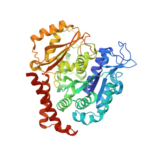
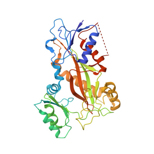
    6D88

  • PubMed Abstract: 

    Colchicine binding site inhibitors (CBSIs) hold great potential in developing new generations of antimitotic drugs. Unlike existing tubulin inhibitors such as paclitaxel, they are generally much less susceptible to resistance caused by the overexpression of drug efflux pumps. The 3,4,5-trimethoxyphenyl (TMP) moiety is a critical component present in many CBSIs, playing an important role in maintaining suitable molecular conformations of CBSIs and contributing to their high binding affinities to tubulin. Previously reported modifications to the TMP moiety in a variety of scaffolds of CBSIs have usually resulted in reduced antiproliferative potency. We previously reported a potent CBSI, VERU-111, that also contains the TMP moiety. Herein, we report the discovery of a VERU-111 analogue 13f that is significantly more potent than VERU-111. The X-ray crystal structure of 13f in complex with tubulin confirms its direct binding to the colchicine site. In addition, 13f exhibited a strong inhibitory effect on tumor growth in vivo.


  • Organizational Affiliation

    Department of Pharmaceutical Sciences, College of Pharmacy , University of Tennessee Health Science Center , Memphis , Tennessee 38163 , United States.


Macromolecules
Find similar proteins by:  (by identity cutoff)  |  3D Structure
Entity ID: 1
MoleculeChains Sequence LengthOrganismDetailsImage
Tubulin alpha-1B chain
A, C
450Sus scrofaMutation(s): 0 
UniProt
Find proteins for Q2XVP4 (Sus scrofa)
Explore Q2XVP4 
Go to UniProtKB:  Q2XVP4
Entity Groups  
Sequence Clusters30% Identity50% Identity70% Identity90% Identity95% Identity100% Identity
UniProt GroupQ2XVP4
Sequence Annotations
Expand
  • Reference Sequence
Find similar proteins by:  (by identity cutoff)  |  3D Structure
Entity ID: 2
MoleculeChains Sequence LengthOrganismDetailsImage
Tubulin beta chain
B, D
445Sus scrofaMutation(s): 0 
UniProt
Find proteins for P02554 (Sus scrofa)
Explore P02554 
Go to UniProtKB:  P02554
Entity Groups  
Sequence Clusters30% Identity50% Identity70% Identity90% Identity95% Identity100% Identity
UniProt GroupP02554
Sequence Annotations
Expand
  • Reference Sequence
Find similar proteins by:  (by identity cutoff)  |  3D Structure
Entity ID: 3
MoleculeChains Sequence LengthOrganismDetailsImage
Stathmin-4143Rattus norvegicusMutation(s): 0 
Gene Names: Stmn4
UniProt
Find proteins for P63043 (Rattus norvegicus)
Explore P63043 
Go to UniProtKB:  P63043
Entity Groups  
Sequence Clusters30% Identity50% Identity70% Identity90% Identity95% Identity100% Identity
UniProt GroupP63043
Sequence Annotations
Expand
  • Reference Sequence
Find similar proteins by:  (by identity cutoff)  |  3D Structure
Entity ID: 4
MoleculeChains Sequence LengthOrganismDetailsImage
Tubulin tyrosine ligase384Gallus gallusMutation(s): 0 
Gene Names: TTL
Entity Groups  
Sequence Clusters30% Identity50% Identity70% Identity90% Identity95% Identity100% Identity
Sequence Annotations
Expand
  • Reference Sequence
Small Molecules
Ligands 7 Unique
IDChains Name / Formula / InChI Key2D Diagram3D Interactions
GTP
Query on GTP

Download Ideal Coordinates CCD File 
G [auth A],
O [auth C]
GUANOSINE-5'-TRIPHOSPHATE
C10 H16 N5 O14 P3
XKMLYUALXHKNFT-UUOKFMHZSA-N
ACP
Query on ACP

Download Ideal Coordinates CCD File 
T [auth F]PHOSPHOMETHYLPHOSPHONIC ACID ADENYLATE ESTER
C11 H18 N5 O12 P3
UFZTZBNSLXELAL-IOSLPCCCSA-N
GDP
Query on GDP

Download Ideal Coordinates CCD File 
J [auth B],
R [auth D]
GUANOSINE-5'-DIPHOSPHATE
C10 H15 N5 O11 P2
QGWNDRXFNXRZMB-UUOKFMHZSA-N
G9K (Subject of Investigation/LOI)
Query on G9K

Download Ideal Coordinates CCD File 
N [auth B],
S [auth D]
[2-(1H-indol-3-yl)-1H-imidazol-4-yl](8-methoxy-1,4-benzodioxin-6-yl)methanone
C21 H15 N3 O4
MUEAWGZGNIDGNE-UHFFFAOYSA-N
MES
Query on MES

Download Ideal Coordinates CCD File 
K [auth B],
L [auth B]
2-(N-MORPHOLINO)-ETHANESULFONIC ACID
C6 H13 N O4 S
SXGZJKUKBWWHRA-UHFFFAOYSA-N
CA
Query on CA

Download Ideal Coordinates CCD File 
H [auth A],
P [auth C]
CALCIUM ION
Ca
BHPQYMZQTOCNFJ-UHFFFAOYSA-N
MG
Query on MG

Download Ideal Coordinates CCD File 
I [auth A],
M [auth B],
Q [auth C]
MAGNESIUM ION
Mg
JLVVSXFLKOJNIY-UHFFFAOYSA-N
Experimental Data & Validation

Experimental Data

  • Method: X-RAY DIFFRACTION
  • Resolution: 2.85 Å
  • R-Value Free: 0.246 
  • R-Value Work: 0.192 
  • R-Value Observed: 0.195 
  • Space Group: P 21 21 21
Unit Cell:
Length ( Å )Angle ( ˚ )
a = 105.536α = 90
b = 157.894β = 90
c = 182.025γ = 90
Software Package:
Software NamePurpose
HKL-2000data scaling
PHENIXrefinement
PDB_EXTRACTdata extraction
HKL-2000data reduction
PHASERphasing

Structure Validation

View Full Validation Report


Ligand Structure Quality Assessment 


Entry History & Funding Information

Deposition Data


Funding OrganizationLocationGrant Number
National Institutes of Health/National Cancer Institute (NIH/NCI)United StatesR01CA148706

Revision History  (Full details and data files)

  • Version 1.0: 2018-09-12
    Type: Initial release
  • Version 1.1: 2018-09-26
    Changes: Data collection, Database references
  • Version 1.2: 2019-12-04
    Changes: Author supporting evidence
  • Version 1.3: 2024-03-13
    Changes: Data collection, Database references, Derived calculations, Refinement description