6D8C

Cryo-EM structure of FLNaABD E254K bound to phalloidin-stabilized F-actin


Experimental Data Snapshot

  • Method: ELECTRON MICROSCOPY
  • Resolution: 3.54 Å
  • Aggregation State: FILAMENT 
  • Reconstruction Method: HELICAL 

wwPDB Validation   3D Report Full Report


This is version 2.0 of the entry. See complete history


Literature

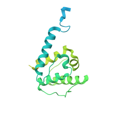
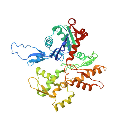
Structural basis of the filamin A actin-binding domain interaction with F-actin.

Iwamoto, D.V.Huehn, A.Simon, B.Huet-Calderwood, C.Baldassarre, M.Sindelar, C.V.Calderwood, D.A.

(2018) Nat Struct Mol Biol 25: 918-927

  • DOI: https://doi.org/10.1038/s41594-018-0128-3
  • Primary Citation of Related Structures:  
    6D8C

  • PubMed Abstract: 

    Actin-cross-linking proteins assemble actin filaments into higher-order structures essential for orchestrating cell shape, adhesion, and motility. Missense mutations in the tandem calponin homology domains of their actin-binding domains (ABDs) underlie numerous genetic diseases, but a molecular understanding of these pathologies is hampered by the lack of high-resolution structures of any actin-cross-linking protein bound to F-actin. Here, taking advantage of a high-affinity, disease-associated mutant of the human filamin A (FLNa) ABD, we combine cryo-electron microscopy and functional studies to reveal at near-atomic resolution how the first calponin homology domain (CH1) and residues immediately N-terminal to it engage actin. We further show that reorientation of CH2 relative to CH1 is required to avoid clashes with actin and to expose F-actin-binding residues on CH1. Our data explain localization of disease-associated loss-of-function mutations to FLNaCH1 and gain-of-function mutations to the regulatory FLNaCH2. Sequence conservation argues that this provides a general model for ABD-F-actin binding.


  • Organizational Affiliation

    Department of Pharmacology, Yale University, New Haven, CT, USA.


Macromolecules
Find similar proteins by:  (by identity cutoff)  |  3D Structure
Entity ID: 1
MoleculeChains Sequence LengthOrganismDetailsImage
Filamin-AA [auth D],
C [auth A],
E [auth B],
G [auth C],
I [auth E]
306Homo sapiensMutation(s): 1 
Gene Names: FLNAFLNFLN1
UniProt & NIH Common Fund Data Resources
Find proteins for P21333 (Homo sapiens)
Explore P21333 
Go to UniProtKB:  P21333
PHAROS:  P21333
GTEx:  ENSG00000196924 
Entity Groups  
Sequence Clusters30% Identity50% Identity70% Identity90% Identity95% Identity100% Identity
UniProt GroupP21333
Sequence Annotations
Expand
  • Reference Sequence
Find similar proteins by:  (by identity cutoff)  |  3D Structure
Entity ID: 2
MoleculeChains Sequence LengthOrganismDetailsImage
Actin, alpha skeletal muscleB [auth H],
D [auth J],
F [auth K],
H [auth L],
J [auth M]
375Gallus gallusMutation(s): 0 
UniProt
Find proteins for P68139 (Gallus gallus)
Explore P68139 
Go to UniProtKB:  P68139
Entity Groups  
Sequence Clusters30% Identity50% Identity70% Identity90% Identity95% Identity100% Identity
UniProt GroupP68139
Sequence Annotations
Expand
  • Reference Sequence

Find similar proteins by:  Sequence   |   3D Structure  

Entity ID: 3
MoleculeChains Sequence LengthOrganismDetailsImage
PhalloidinK [auth N],
L [auth O],
M [auth P]
7Amanita phalloidesMutation(s): 0 
UniProt
Find proteins for P0CU65 (Amanita phalloides)
Explore P0CU65 
Go to UniProtKB:  P0CU65
Entity Groups  
UniProt GroupP0CU65
Sequence Annotations
Expand
  • Reference Sequence
Small Molecules
Ligands 2 Unique
IDChains Name / Formula / InChI Key2D Diagram3D Interactions
ADP
Query on ADP

Download Ideal Coordinates CCD File 
N [auth H],
P [auth J],
R [auth K],
T [auth L],
V [auth M]
ADENOSINE-5'-DIPHOSPHATE
C10 H15 N5 O10 P2
XTWYTFMLZFPYCI-KQYNXXCUSA-N
MG
Query on MG

Download Ideal Coordinates CCD File 
O [auth H],
Q [auth J],
S [auth K],
U [auth L],
W [auth M]
MAGNESIUM ION
Mg
JLVVSXFLKOJNIY-UHFFFAOYSA-N
Modified Residues  3 Unique
IDChains TypeFormula2D DiagramParent
ALO
Query on ALO
K [auth N],
L [auth O],
M [auth P]
L-PEPTIDE LINKINGC4 H9 N O3THR
G5G
Query on G5G
K [auth N],
L [auth O],
M [auth P]
L-PEPTIDE LINKINGC6 H13 N O4LEU
HYP
Query on HYP
K [auth N],
L [auth O],
M [auth P]
L-PEPTIDE LINKINGC5 H9 N O3PRO
Biologically Interesting Molecules (External Reference) 1 Unique
Experimental Data & Validation

Experimental Data

  • Method: ELECTRON MICROSCOPY
  • Resolution: 3.54 Å
  • Aggregation State: FILAMENT 
  • Reconstruction Method: HELICAL 
EM Software:
TaskSoftware PackageVersion
MODEL REFINEMENTPHENIX
RECONSTRUCTIONRELION2.0.3

Structure Validation

View Full Validation Report


Entry History 

Deposition Data

Revision History  (Full details and data files)

  • Version 1.0: 2018-09-19
    Type: Initial release
  • Version 1.1: 2018-10-03
    Changes: Data collection, Database references
  • Version 1.2: 2018-10-17
    Changes: Data collection, Database references
  • Version 2.0: 2023-11-15
    Changes: Atomic model, Data collection, Database references, Derived calculations, Refinement description