6DI4

Rational Modification of Vanillin Derivatives to Stereospecifically Destabilize Sickle Hemoglobin Polymer Formation

  • Classification: OXYGEN TRANSPORT
  • Organism(s): Homo sapiens
  • Mutation(s): No 

  • Deposited: 2018-05-22 Released: 2018-09-05 
  • Deposition Author(s): Pagare, P.P., Musayev, F.N.
  • Funding Organization(s): National Institutes of Health/National Institute on Minority Health and Health Disparities (NIH/NIMHD), National Institutes of Health/National Center for Research Resources (NIH/NCRR)

Experimental Data Snapshot

  • Method: X-RAY DIFFRACTION
  • Resolution: 1.90 Å
  • R-Value Free: 0.249 
  • R-Value Work: 0.195 
  • R-Value Observed: 0.195 

wwPDB Validation   3D Report Full Report


Ligand Structure Quality Assessment 


This is version 1.2 of the entry. See complete history


Literature

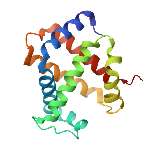
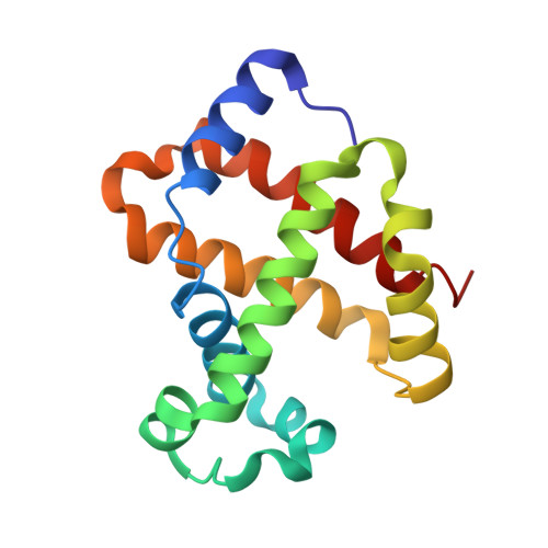
Rational modification of vanillin derivatives to stereospecifically destabilize sickle hemoglobin polymer formation.

Deshpande, T.M.Pagare, P.P.Ghatge, M.S.Chen, Q.Musayev, F.N.Venitz, J.Zhang, Y.Abdulmalik, O.Safo, M.K.

(2018) Acta Crystallogr D Struct Biol 74: 956-964

  • DOI: https://doi.org/10.1107/S2059798318009919
  • Primary Citation of Related Structures:  
    6DI4

  • PubMed Abstract: 

    Increasing the affinity of hemoglobin for oxygen represents a feasible and promising therapeutic approach for sickle cell disease by mitigating the primary pathophysiological event, i.e. the hypoxia-induced polymerization of sickle hemoglobin (Hb S) and the concomitant erythrocyte sickling. Investigations on a novel synthetic antisickling agent, SAJ-310, with improved and sustained antisickling activity have previously been reported. To further enhance the biological effects of SAJ-310, a structure-based approach was employed to modify this compound to specifically inhibit Hb S polymer formation through interactions which perturb the Hb S polymer-stabilizing αF-helix, in addition to primarily increasing the oxygen affinity of hemoglobin. Three compounds, TD-7, TD-8 and TD-9, were synthesized and studied for their interactions with hemoglobin at the atomic level, as well as their functional and antisickling activities in vitro. X-ray crystallographic studies with liganded hemoglobin in complex with TD-7 showed the predicted mode of binding, although the interaction with the αF-helix was not as strong as expected. These findings provide important insights and guidance towards the development of molecules that would be expected to bind and make stronger interactions with the αF-helix, resulting in more efficacious novel therapeutics for sickle cell disease.


  • Organizational Affiliation

    Department of Medicinal Chemistry, School of Pharmacy, Virginia Commonwealth University, 800 East Leigh Street, Richmond, VA 23219, USA.


Macromolecules
Find similar proteins by:  (by identity cutoff)  |  3D Structure
Entity ID: 1
MoleculeChains Sequence LengthOrganismDetailsImage
Hemoglobin subunit alpha
A, C
141Homo sapiensMutation(s): 0 
UniProt & NIH Common Fund Data Resources
Find proteins for P69905 (Homo sapiens)
Explore P69905 
Go to UniProtKB:  P69905
PHAROS:  P69905
Entity Groups  
Sequence Clusters30% Identity50% Identity70% Identity90% Identity95% Identity100% Identity
UniProt GroupP69905
Sequence Annotations
Expand
  • Reference Sequence
Find similar proteins by:  (by identity cutoff)  |  3D Structure
Entity ID: 2
MoleculeChains Sequence LengthOrganismDetailsImage
Hemoglobin subunit beta
B, D
146Homo sapiensMutation(s): 0 
UniProt & NIH Common Fund Data Resources
Find proteins for P68871 (Homo sapiens)
Explore P68871 
Go to UniProtKB:  P68871
PHAROS:  P68871
GTEx:  ENSG00000244734 
Entity Groups  
Sequence Clusters30% Identity50% Identity70% Identity90% Identity95% Identity100% Identity
UniProt GroupP68871
Sequence Annotations
Expand
  • Reference Sequence
Small Molecules
Ligands 3 Unique
IDChains Name / Formula / InChI Key2D Diagram3D Interactions
HEM
Query on HEM

Download Ideal Coordinates CCD File 
F [auth A],
I [auth B],
K [auth C],
N [auth D]
PROTOPORPHYRIN IX CONTAINING FE
C34 H32 Fe N4 O4
KABFMIBPWCXCRK-RGGAHWMASA-L
GJ1
Query on GJ1

Download Ideal Coordinates CCD File 
G [auth A],
L [auth C]
{6-[(4-methoxy-2-methylphenoxy)methyl]pyridin-2-yl}methanol
C15 H17 N O3
GBTFLQPUSKDFPL-UHFFFAOYSA-N
CMO
Query on CMO

Download Ideal Coordinates CCD File 
E [auth A],
H [auth B],
J [auth C],
M [auth D]
CARBON MONOXIDE
C O
UGFAIRIUMAVXCW-UHFFFAOYSA-N
Experimental Data & Validation

Experimental Data

  • Method: X-RAY DIFFRACTION
  • Resolution: 1.90 Å
  • R-Value Free: 0.249 
  • R-Value Work: 0.195 
  • R-Value Observed: 0.195 
  • Space Group: P 21 21 21
Unit Cell:
Length ( Å )Angle ( ˚ )
a = 62.696α = 90
b = 83.404β = 90
c = 104.934γ = 90
Software Package:
Software NamePurpose
CNSrefinement
CrystalCleardata scaling
CrystalCleardata collection
CrysalisProdata reduction
PHENIXmodel building

Structure Validation

View Full Validation Report


Ligand Structure Quality Assessment 


Entry History & Funding Information

Deposition Data


Funding OrganizationLocationGrant Number
National Institutes of Health/National Institute on Minority Health and Health Disparities (NIH/NIMHD)United StatesMD009124
National Institutes of Health/National Center for Research Resources (NIH/NCRR)United StatesS10-OD021756

Revision History  (Full details and data files)

  • Version 1.0: 2018-09-05
    Type: Initial release
  • Version 1.1: 2018-10-17
    Changes: Data collection, Database references
  • Version 1.2: 2019-12-04
    Changes: Author supporting evidence