6E21

Joint X-ray/neutron structure of PKAc with products Sr2-ADP and phosphorylated peptide SP20


Experimental Data Snapshot

  • Method: X-RAY DIFFRACTION
  • Resolution: 2.00 Å
  • R-Value Free: 0.217 
  • R-Value Work: 0.187 

  • Method: NEUTRON DIFFRACTION
  • Resolution: 2.50 Å
  • R-Value Free: 0.275 
  • R-Value Work: 0.226 

wwPDB Validation   3D Report Full Report


This is version 1.2 of the entry. See complete history


Literature

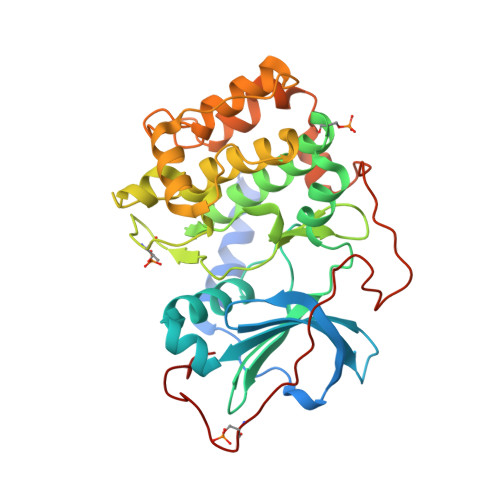
Zooming in on protons: Neutron structure of protein kinase A trapped in a product complex.

Gerlits, O.Weiss, K.L.Blakeley, M.P.Veglia, G.Taylor, S.S.Kovalevsky, A.

(2019) Sci Adv 5: eaav0482-eaav0482

  • DOI: https://doi.org/10.1126/sciadv.aav0482
  • Primary Citation of Related Structures:  
    6E21

  • PubMed Abstract: 

    The question vis-à-vis the chemistry of phosphoryl group transfer catalyzed by protein kinases remains a major challenge. The neutron diffraction structure of the catalytic subunit of cAMP-dependent protein kinase (PKA-C) provides a more complete chemical portrait of key proton interactions at the active site. By using a high-affinity protein kinase substrate (PKS) peptide, we captured the reaction products, dephosphorylated nucleotide [adenosine diphosphate (ADP)] and phosphorylated PKS (pPKS), bound at the active site. In the complex, the phosphoryl group of the peptide is protonated, whereas the carboxyl group of the catalytic Asp 166 is not. Our structure, including conserved waters, shows how the peptide links the distal parts of the cleft together, creating a network that engages the entire molecule. By comparing slow-exchanging backbone amides to those determined by the NMR analysis of PKA-C with ADP and inhibitor peptide (PKI), we identified exchangeable amides that likely distinguish catalytic and inhibited states.


  • Organizational Affiliation

    Bredesen Center, University of Tennessee, Knoxville, TN 37996, USA.


Macromolecules
Find similar proteins by:  (by identity cutoff)  |  3D Structure
Entity ID: 1
MoleculeChains Sequence LengthOrganismDetailsImage
cAMP-dependent protein kinase catalytic subunit alpha356Mus musculusMutation(s): 0 
Gene Names: PrkacaPkaca
EC: 2.7.11.11
UniProt
Find proteins for P05132 (Mus musculus)
Explore P05132 
Go to UniProtKB:  P05132
Entity Groups  
Sequence Clusters30% Identity50% Identity70% Identity90% Identity95% Identity100% Identity
UniProt GroupP05132
Sequence Annotations
Expand
  • Reference Sequence

Find similar proteins by:  Sequence   |   3D Structure  

Entity ID: 2
MoleculeChains Sequence LengthOrganismDetailsImage
pSP2020Homo sapiensMutation(s): 0 
UniProt & NIH Common Fund Data Resources
Find proteins for P61925 (Homo sapiens)
Explore P61925 
Go to UniProtKB:  P61925
PHAROS:  P61925
GTEx:  ENSG00000171033 
Entity Groups  
Sequence Clusters30% Identity50% Identity70% Identity90% Identity95% Identity100% Identity
UniProt GroupP61925
Sequence Annotations
Expand
  • Reference Sequence
Small Molecules
Ligands 2 Unique
IDChains Name / Formula / InChI Key2D Diagram3D Interactions
ADP
Query on ADP

Download Ideal Coordinates CCD File 
E [auth A]ADENOSINE-5'-DIPHOSPHATE
C10 H15 N5 O10 P2
XTWYTFMLZFPYCI-KQYNXXCUSA-N
SR
Query on SR

Download Ideal Coordinates CCD File 
C [auth A],
D [auth A]
STRONTIUM ION
Sr
PWYYWQHXAPXYMF-UHFFFAOYSA-N
Modified Residues  2 Unique
IDChains TypeFormula2D DiagramParent
SEP
Query on SEP
A
L-PEPTIDE LINKINGC3 H8 N O6 PSER
TPO
Query on TPO
A
L-PEPTIDE LINKINGC4 H10 N O6 PTHR
Experimental Data & Validation

Experimental Data

  • Method: X-RAY DIFFRACTION
  • Resolution: 2.00 Å
  • R-Value Free: 0.217 
  • R-Value Work: 0.187 
  • Space Group: P 21 21 21
  • Method: NEUTRON DIFFRACTION
  • Resolution: 2.50 Å
  • R-Value Free: 0.275 
  • R-Value Work: 0.226 
  • Space Group: P 21 21 21
Unit Cell:
Length ( Å )Angle ( ˚ )
a = 59.332α = 90
b = 79.898β = 90
c = 100.292γ = 90
Software Package:
Software NamePurpose
nCNSrefinement
HKL-3000data reduction
HKL-3000data scaling
PHASERphasing
LAUEGENdata reduction
LSCALEdata scaling
CNSphasing

Structure Validation

View Full Validation Report


Entry History 

Deposition Data

Revision History  (Full details and data files)

  • Version 1.0: 2019-04-03
    Type: Initial release
  • Version 1.1: 2019-04-10
    Changes: Data collection, Database references
  • Version 1.2: 2023-10-25
    Changes: Data collection, Database references, Derived calculations, Refinement description