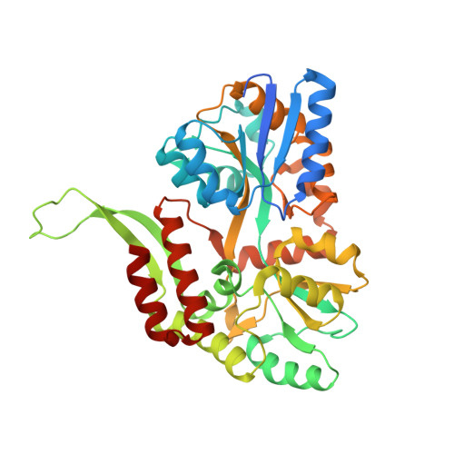
6EQZ

A MamC-MIC insertion in MBP scaffold at position K170


Experimental Data Snapshot

  • Method: X-RAY DIFFRACTION
  • Resolution: 2.29 Å
  • R-Value Free: 0.274 
  • R-Value Work: 0.205 
  • R-Value Observed: 0.208 

wwPDB Validation   3D Report Full Report


This is version 2.1 of the entry. See complete history


Literature

The importance of the helical structure of a MamC-derived magnetite-interacting peptide for its function in magnetite formation.

Nudelman, H.Perez Gonzalez, T.Kolushiva, S.Widdrat, M.Reichel, V.Peigneux, A.Davidov, G.Bitton, R.Faivre, D.Jimenez-Lopez, C.Zarivach, R.

(2018) Acta Crystallogr D Struct Biol 74: 10-20

  • DOI: https://doi.org/10.1107/S2059798317017491
  • Primary Citation of Related Structures:  
    5MM3, 6EQZ

  • PubMed Abstract: 

    Biomineralization is the process of mineral formation by organisms and involves the uptake of ions from the environment in order to produce minerals, with the process generally being mediated by proteins. Most proteins that are involved in mineral interactions are predicted to contain disordered regions containing large numbers of negatively charged amino acids. Magnetotactic bacteria, which are used as a model system for iron biomineralization, are Gram-negative bacteria that can navigate through geomagnetic fields using a specific organelle, the magnetosome. Each organelle comprises a membrane-enveloped magnetic nanoparticle, magnetite, the formation of which is controlled by a specific set of proteins. One of the most abundant of these proteins is MamC, a small magnetosome-associated integral membrane protein that contains two transmembrane α-helices connected by an ∼21-amino-acid peptide. In vitro studies of this MamC peptide showed that it forms a helical structure that can interact with the magnetite surface and affect the size and shape of the growing crystal. Our results show that a disordered structure of the MamC magnetite-interacting component (MamC-MIC) abolishes its interaction with magnetite particles. Moreover, the size and shape of magnetite crystals grown in in vitro magnetite-precipitation experiments in the presence of this disordered peptide were different from the traits of crystals grown in the presence of other peptides or in the presence of the helical MIC. It is suggested that the helical structure of the MamC-MIC is important for its function during magnetite formation.


  • Organizational Affiliation

    Department of Life Sciences and the National Institute for Biotechnology in the Negev, Ben-Gurion University of the Negev, Beer Sheva, Israel.


Macromolecules
Find similar proteins by:  (by identity cutoff)  |  3D Structure
Entity ID: 1
MoleculeChains Sequence LengthOrganismDetailsImage
Maltose-binding periplasmic protein,Tightly bound bacterial magnetic particle protein,Maltose-binding periplasmic proteinA,
B,
C [auth D],
D [auth G]
401Escherichia coli O157:H7Paramagnetospirillum magneticum AMB-1Mutation(s): 0 
Gene Names: malEZ5632ECs5017mms13amb0951
UniProt
Find proteins for P0AEX9 (Escherichia coli (strain K12))
Explore P0AEX9 
Go to UniProtKB:  P0AEX9
Find proteins for Q2W8S0 (Magnetospirillum magneticum (strain AMB-1 / ATCC 700264))
Explore Q2W8S0 
Go to UniProtKB:  Q2W8S0
Entity Groups  
Sequence Clusters30% Identity50% Identity70% Identity90% Identity95% Identity100% Identity
UniProt GroupsQ2W8S0P0AEX9
Sequence Annotations
Expand
  • Reference Sequence
Oligosaccharides

Help

Entity ID: 2
MoleculeChains Length2D Diagram Glycosylation3D Interactions
alpha-D-glucopyranose-(1-4)-alpha-D-glucopyranoseE [auth C],
F [auth E],
G [auth F],
H
2N/A
Glycosylation Resources
GlyTouCan:  G07411ON
GlyCosmos:  G07411ON
Experimental Data & Validation

Experimental Data

  • Method: X-RAY DIFFRACTION
  • Resolution: 2.29 Å
  • R-Value Free: 0.274 
  • R-Value Work: 0.205 
  • R-Value Observed: 0.208 
  • Space Group: P 21 21 21
Unit Cell:
Length ( Å )Angle ( ˚ )
a = 110.682α = 90
b = 113.813β = 90
c = 115.511γ = 90
Software Package:
Software NamePurpose
PHENIXrefinement
XDSdata scaling
REFMACphasing

Structure Validation

View Full Validation Report


Entry History 

Deposition Data

Revision History  (Full details and data files)

  • Version 1.0: 2017-11-01
    Type: Initial release
  • Version 1.1: 2018-01-03
    Changes: Database references
  • Version 1.2: 2018-02-07
    Changes: Database references, Source and taxonomy
  • Version 2.0: 2020-07-29
    Type: Remediation
    Reason: Carbohydrate remediation
    Changes: Atomic model, Data collection, Derived calculations, Non-polymer description, Structure summary
  • Version 2.1: 2024-01-17
    Changes: Data collection, Database references, Refinement description, Structure summary