6FCZ

Model of gC1q-Fc complex based on 7A EM map


Experimental Data Snapshot

  • Method: ELECTRON MICROSCOPY
  • Resolution: 10.0 Å
  • Aggregation State: PARTICLE 
  • Reconstruction Method: SINGLE PARTICLE 

wwPDB Validation   3D Report Full Report


This is version 1.1 of the entry. See complete history


Literature

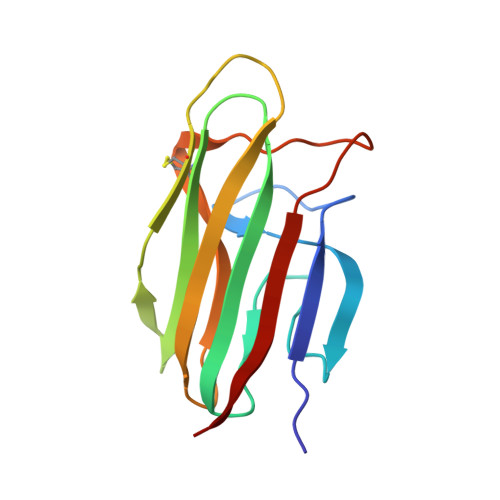
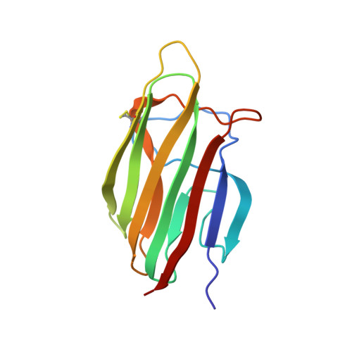
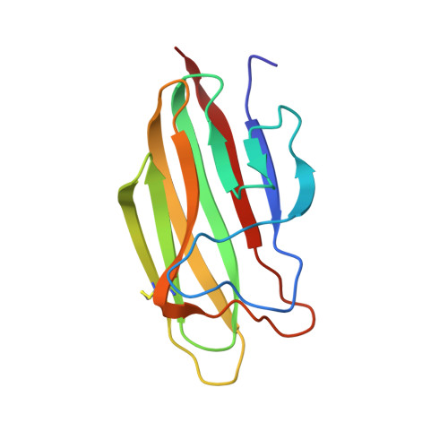
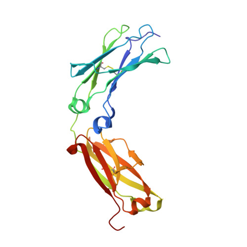
Structures of C1-IgG1 provide insights into how danger pattern recognition activates complement.

Ugurlar, D.Howes, S.C.de Kreuk, B.J.Koning, R.I.de Jong, R.N.Beurskens, F.J.Schuurman, J.Koster, A.J.Sharp, T.H.Parren, P.W.H.I.Gros, P.

(2018) Science 359: 794-797

  • DOI: https://doi.org/10.1126/science.aao4988
  • Primary Citation of Related Structures:  
    6FCZ

  • PubMed Abstract: 

    Danger patterns on microbes or damaged host cells bind and activate C1, inducing innate immune responses and clearance through the complement cascade. How these patterns trigger complement initiation remains elusive. Here, we present cryo-electron microscopy analyses of C1 bound to monoclonal antibodies in which we observed heterogeneous structures of single and clustered C1-immunoglobulin G1 (IgG1) hexamer complexes. Distinct C1q binding sites are observed on the two Fc-CH2 domains of each IgG molecule. These are consistent with known interactions and also reveal additional interactions, which are supported by functional IgG1-mutant analysis. Upon antibody binding, the C1q arms condense, inducing rearrangements of the C1r 2 s 2 proteases and tilting C1q's cone-shaped stalk. The data suggest that C1r may activate C1s within single, strained C1 complexes or between neighboring C1 complexes on surfaces.


  • Organizational Affiliation

    Crystal and Structural Chemistry, Bijvoet Center for Biomolecular Research, Department of Chemistry, Faculty of Science, Utrecht University, Padualaan 8, 3584 CH Utrecht, Netherlands.


Macromolecules
Find similar proteins by:  (by identity cutoff)  |  3D Structure
Entity ID: 1
MoleculeChains Sequence LengthOrganismDetailsImage
Complement C1q subcomponent subunit A133Homo sapiensMutation(s): 0 
UniProt & NIH Common Fund Data Resources
Find proteins for P02745 (Homo sapiens)
Explore P02745 
Go to UniProtKB:  P02745
PHAROS:  P02745
GTEx:  ENSG00000173372 
Entity Groups  
Sequence Clusters30% Identity50% Identity70% Identity90% Identity95% Identity100% Identity
UniProt GroupP02745
Sequence Annotations
Expand
  • Reference Sequence
Find similar proteins by:  (by identity cutoff)  |  3D Structure
Entity ID: 2
MoleculeChains Sequence LengthOrganismDetailsImage
Complement C1q subcomponent subunit B132Homo sapiensMutation(s): 0 
UniProt & NIH Common Fund Data Resources
Find proteins for P02746 (Homo sapiens)
Explore P02746 
Go to UniProtKB:  P02746
PHAROS:  P02746
Entity Groups  
Sequence Clusters30% Identity50% Identity70% Identity90% Identity95% Identity100% Identity
UniProt GroupP02746
Sequence Annotations
Expand
  • Reference Sequence
Find similar proteins by:  (by identity cutoff)  |  3D Structure
Entity ID: 3
MoleculeChains Sequence LengthOrganismDetailsImage
Complement C1q subcomponent subunit C129Homo sapiensMutation(s): 0 
UniProt & NIH Common Fund Data Resources
Find proteins for P02747 (Homo sapiens)
Explore P02747 
Go to UniProtKB:  P02747
PHAROS:  P02747
GTEx:  ENSG00000159189 
Entity Groups  
Sequence Clusters30% Identity50% Identity70% Identity90% Identity95% Identity100% Identity
UniProt GroupP02747
Sequence Annotations
Expand
  • Reference Sequence
Find similar proteins by:  (by identity cutoff)  |  3D Structure
Entity ID: 4
MoleculeChains Sequence LengthOrganismDetailsImage
Immunoglobulin gamma-1 heavy chainD [auth H],
E [auth K]
216Homo sapiensMutation(s): 0 
UniProt
Find proteins for P0DOX5 (Homo sapiens)
Explore P0DOX5 
Go to UniProtKB:  P0DOX5
Entity Groups  
Sequence Clusters30% Identity50% Identity70% Identity90% Identity95% Identity100% Identity
UniProt GroupP0DOX5
Sequence Annotations
Expand
  • Reference Sequence
Experimental Data & Validation

Experimental Data

  • Method: ELECTRON MICROSCOPY
  • Resolution: 10.0 Å
  • Aggregation State: PARTICLE 
  • Reconstruction Method: SINGLE PARTICLE 

Structure Validation

View Full Validation Report


Entry History & Funding Information

Deposition Data


Funding OrganizationLocationGrant Number
Netherlands Organisation for Scientific ResearchNetherlandsCW 714.013.002

Revision History  (Full details and data files)

  • Version 1.0: 2018-02-28
    Type: Initial release
  • Version 1.1: 2018-10-24
    Changes: Advisory, Data collection, Derived calculations