6FQF

THE X-RAY STRUCTURE OF FERRIC-BETA4 HUMAN HEMOGLOBIN


Experimental Data Snapshot

  • Method: X-RAY DIFFRACTION
  • Resolution: 2.10 Å
  • R-Value Free: 0.265 
  • R-Value Work: 0.204 
  • R-Value Observed: 0.207 

wwPDB Validation   3D Report Full Report


Ligand Structure Quality Assessment 


This is version 1.1 of the entry. See complete history


Literature

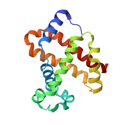
Crystal structure of the ferric homotetrameric beta4human hemoglobin.

Mazzarella, L.Merlino, A.Balasco, N.Balsamo, A.Vergara, A.

(2018) Biophys Chem 240: 9-14

  • DOI: https://doi.org/10.1016/j.bpc.2018.05.003
  • Primary Citation of Related Structures:  
    6FQF

  • PubMed Abstract: 

    Spectroscopic studies carried out in the early seventies have shown that the β-homotetramer of human hemoglobin (β 4 -HbA) in the ferric state is a mixture of aquomet and bis-histidyl forms. Here we present the first crystal structure, solved at 2.10 Å resolution, of the oxidized form of β 4 -HbA. The overall quaternary structure of the protein in the ferric state is virtually indistinguishable from that of the ferrous deoxygenated and carbomonoxy forms. The structure reveals that the four hemes are exclusively in an aquomet coordination, without any trace of bis-histidyl coordination. The oxidation of β 4 -HbA is associated with the formation of a disulfide bridge between residues Cys112(G14) of β 1 4 and β 2 3 chains. The coordination state of β 4 -HbA has been compared to that known for other organisms that exhibit bis-histidyl heme coordination in the β 4 state. This occurrence has been discussed in terms of different organism physiology.


  • Organizational Affiliation

    Department of Chemical Sciences, University of Naples "Federico II", Naples, Italy; Accademia di Scienze Fisiche e Matematiche della Società Nazionale di Scienze, Lettere ed Arti in Napoli, Naples, Italy.


Macromolecules
Find similar proteins by:  (by identity cutoff)  |  3D Structure
Entity ID: 1
MoleculeChains Sequence LengthOrganismDetailsImage
Hemoglobin subunit beta
A, B, C, D
146Homo sapiensMutation(s): 0 
Gene Names: HBB
UniProt & NIH Common Fund Data Resources
Find proteins for P68871 (Homo sapiens)
Explore P68871 
Go to UniProtKB:  P68871
PHAROS:  P68871
GTEx:  ENSG00000244734 
Entity Groups  
Sequence Clusters30% Identity50% Identity70% Identity90% Identity95% Identity100% Identity
UniProt GroupP68871
Sequence Annotations
Expand
  • Reference Sequence
Experimental Data & Validation

Experimental Data

  • Method: X-RAY DIFFRACTION
  • Resolution: 2.10 Å
  • R-Value Free: 0.265 
  • R-Value Work: 0.204 
  • R-Value Observed: 0.207 
  • Space Group: P 1 21 1
Unit Cell:
Length ( Å )Angle ( ˚ )
a = 52.7α = 90
b = 82.1β = 91.3
c = 62.5γ = 90
Software Package:
Software NamePurpose
REFMACrefinement
HKL-2000data reduction
HKL-2000data scaling
AMoREphasing

Structure Validation

View Full Validation Report


Ligand Structure Quality Assessment 


Entry History 

Deposition Data

Revision History  (Full details and data files)

  • Version 1.0: 2018-06-13
    Type: Initial release
  • Version 1.1: 2024-01-17
    Changes: Data collection, Database references, Derived calculations, Refinement description