6H01

Crystal structure of a domain-swapped dark-state sfGFP containing the unnatural amino acid ortho-nitrobenzyl-tyrosine (ONBY) at residue 66


Experimental Data Snapshot

  • Method: X-RAY DIFFRACTION
  • Resolution: 2.78 Å
  • R-Value Free: 0.239 
  • R-Value Work: 0.201 
  • R-Value Observed: 0.203 

wwPDB Validation   3D Report Full Report


This is version 1.2 of the entry. See complete history


Literature

Crystal structure of a domain-swapped photoactivatable sfGFP variant provides evidence for GFP folding pathway.

Kesgin-Schaefer, S.Heidemann, J.Puchert, A.Koelbel, K.Yorke, B.A.Huse, N.Pearson, A.R.Uetrecht, C.Tidow, H.

(2019) FEBS J 286: 2329-2340

  • DOI: https://doi.org/10.1111/febs.14797
  • Primary Citation of Related Structures:  
    6H01

  • PubMed Abstract: 

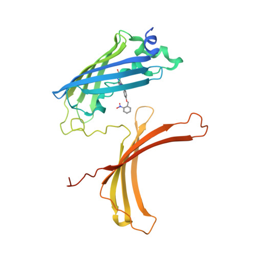
    Photoactivatable fluorescent proteins (PA-FPs) are a powerful non-invasive tool in high-resolution live-cell imaging. They can be converted from an inactive to an active form by light, enabling the spatial and temporal trafficking of proteins and cell dynamics. PA-FPs have been previously generated by mutating selected residues in the chromophore or in its close proximity. A new strategy to generate PA-FPs is the genetic incorporation of unnatural amino acids (UAAs) containing photocaged groups using unique suppressor tRNA/aminoacyl-tRNA synthetase pairs. We set out to develop a photoactivatable GFP variant suitable for time-resolved structural studies. Here, we report the crystal structure of superfolder GFP (sfGFP) containing the UAA ortho-nitrobenzyl-tyrosine (ONBY) at position 66 and its spectroscopic characterization. Surprisingly, the crystal structure (to 2.7 Å resolution) reveals a dimeric domain-swapped arrangement of sfGFP66ONBY with residues 1-142 of one molecule associating with residues 148-234 from another molecule. This unusual domain-swapped structure supports a previously postulated GFP folding pathway that proceeds via an equilibrium intermediate.


  • Organizational Affiliation

    The Hamburg Centre for Ultrafast Imaging, Germany.


Macromolecules
Find similar proteins by:  (by identity cutoff)  |  3D Structure
Entity ID: 1
MoleculeChains Sequence LengthOrganismDetailsImage
Green fluorescent protein
A, B, C, D
264Aequorea victoriaMutation(s): 1 
Gene Names: gfp
UniProt
Find proteins for A0A059PIQ0 (Aequorea victoria)
Explore A0A059PIQ0 
Go to UniProtKB:  A0A059PIQ0
Entity Groups  
Sequence Clusters30% Identity50% Identity70% Identity90% Identity95% Identity100% Identity
UniProt GroupA0A059PIQ0
Sequence Annotations
Expand
  • Reference Sequence
Small Molecules
Modified Residues  1 Unique
IDChains TypeFormula2D DiagramParent
FHE
Query on FHE
A, B, C, D
L-PEPTIDE LINKINGC22 H20 N4 O7THR, TYR, GLY
Experimental Data & Validation

Experimental Data

  • Method: X-RAY DIFFRACTION
  • Resolution: 2.78 Å
  • R-Value Free: 0.239 
  • R-Value Work: 0.201 
  • R-Value Observed: 0.203 
  • Space Group: P 41 21 2
Unit Cell:
Length ( Å )Angle ( ˚ )
a = 155.324α = 90
b = 155.324β = 90
c = 161.977γ = 90
Software Package:
Software NamePurpose
PHENIXrefinement
DIALSdata reduction
Aimlessdata scaling
PHENIXphasing

Structure Validation

View Full Validation Report


Entry History & Funding Information

Deposition Data


Funding OrganizationLocationGrant Number
German Research FoundationGermanyDFG EXC 1074

Revision History  (Full details and data files)

  • Version 1.0: 2019-04-24
    Type: Initial release
  • Version 1.1: 2019-06-26
    Changes: Data collection, Database references
  • Version 1.2: 2024-01-17
    Changes: Data collection, Database references, Derived calculations, Refinement description