6HT2

STRUCTURE OF HEWL BY ELECTRON DIFFRACTION AND MICROFOCUS DIFFRACTION


Experimental Data Snapshot

  • Method: X-RAY DIFFRACTION
  • Resolution: 2.60 Å
  • R-Value Free: 0.252 
  • R-Value Work: 0.199 
  • R-Value Observed: 0.201 

wwPDB Validation   3D Report Full Report


This is version 1.2 of the entry. See complete history


Literature

Nanobeam precession-assisted 3D electron diffraction reveals a new polymorph of hen egg-white lysozyme.

Lanza, A.Margheritis, E.Mugnaioli, E.Cappello, V.Garau, G.Gemmi, M.

(2019) IUCrJ 6: 178-188

  • DOI: https://doi.org/10.1107/S2052252518017657
  • Primary Citation of Related Structures:  
    6HT2, 6HU5

  • PubMed Abstract: 

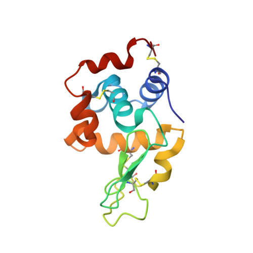
    Recent advances in 3D electron diffraction have allowed the structure determination of several model proteins from submicrometric crystals, the unit-cell parameters and structures of which could be immediately validated by known models previously obtained by X-ray crystallography. Here, the first new protein structure determined by 3D electron diffraction data is presented: a previously unobserved polymorph of hen egg-white lysozyme. This form, with unit-cell parameters a = 31.9, b = 54.4, c = 71.8 Å, β = 98.8°, grows as needle-shaped submicrometric crystals simply by vapor diffusion starting from previously reported crystallization conditions. Remarkably, the data were collected using a low-dose stepwise experimental setup consisting of a precession-assisted nanobeam of ∼150 nm, which has never previously been applied for solving protein structures. The crystal structure was additionally validated using X-ray synchrotron-radiation sources by both powder diffraction and single-crystal micro-diffraction. 3D electron diffraction can be used for the structural characterization of submicrometric macromolecular crystals and is able to identify novel protein polymorphs that are hardly visible in conventional X-ray diffraction experiments. Additionally, the analysis, which was performed on both nanocrystals and microcrystals from the same crystallization drop, suggests that an integrated view from 3D electron diffraction and X-ray microfocus diffraction can be applied to obtain insights into the molecular dynamics during protein crystal growth.


  • Organizational Affiliation

    Center for Nanotechnology Innovation@NEST, Istituto Italiano di Tecnologia, Piazza San Silvestro 12, 56127 Pisa, Italy.


Macromolecules
Find similar proteins by:  (by identity cutoff)  |  3D Structure
Entity ID: 1
MoleculeChains Sequence LengthOrganismDetailsImage
Lysozyme C
A, B
129Gallus gallusMutation(s): 0 
EC: 3.2.1.17
UniProt
Find proteins for P00698 (Gallus gallus)
Explore P00698 
Go to UniProtKB:  P00698
Entity Groups  
Sequence Clusters30% Identity50% Identity70% Identity90% Identity95% Identity100% Identity
UniProt GroupP00698
Sequence Annotations
Expand
  • Reference Sequence
Small Molecules
Ligands 1 Unique
IDChains Name / Formula / InChI Key2D Diagram3D Interactions
CL
Query on CL

Download Ideal Coordinates CCD File 
C [auth A]
D [auth A]
E [auth A]
F [auth A]
G [auth A]
C [auth A],
D [auth A],
E [auth A],
F [auth A],
G [auth A],
H [auth A],
I [auth A],
J [auth A],
K [auth A],
L [auth A],
M [auth A],
N [auth B],
O [auth B],
P [auth B],
Q [auth B],
R [auth B],
S [auth B],
T [auth B],
U [auth B]
CHLORIDE ION
Cl
VEXZGXHMUGYJMC-UHFFFAOYSA-M
Experimental Data & Validation

Experimental Data

  • Method: X-RAY DIFFRACTION
  • Resolution: 2.60 Å
  • R-Value Free: 0.252 
  • R-Value Work: 0.199 
  • R-Value Observed: 0.201 
  • Space Group: P 1 21 1
Unit Cell:
Length ( Å )Angle ( ˚ )
a = 31.7α = 90
b = 53.5β = 99
c = 71.6γ = 90
Software Package:
Software NamePurpose
XDSdata reduction
Aimlessdata scaling
PHASERphasing
REFMACrefinement

Structure Validation

View Full Validation Report


Entry History 

Deposition Data

  • Released Date: 2019-01-23 
  • Deposition Author(s): Garau, G.

Revision History  (Full details and data files)

  • Version 1.0: 2019-01-23
    Type: Initial release
  • Version 1.1: 2019-03-27
    Changes: Data collection, Database references
  • Version 1.2: 2024-01-24
    Changes: Data collection, Database references, Refinement description