6I04

Crystal structure of Sema domain of the Met receptor in complex with FAB


Experimental Data Snapshot

  • Method: X-RAY DIFFRACTION
  • Resolution: 3.10 Å
  • R-Value Free: 0.261 
  • R-Value Work: 0.202 
  • R-Value Observed: 0.205 

wwPDB Validation   3D Report Full Report


This is version 1.3 of the entry. See complete history


Literature

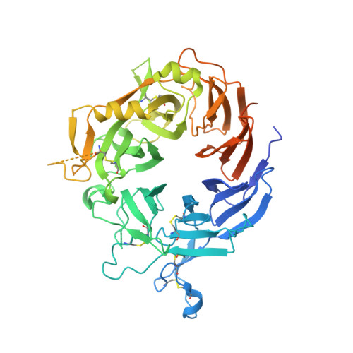
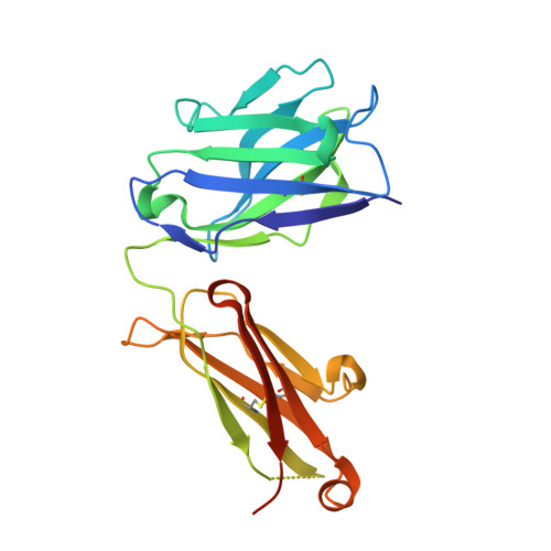
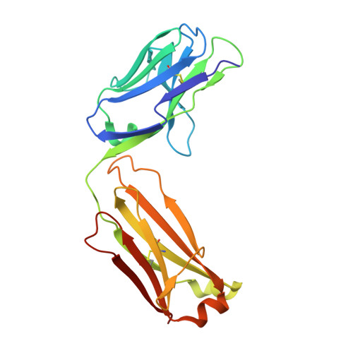
MM-131, a bispecific anti-Met/EpCAM mAb, inhibits HGF-dependent and HGF-independent Met signaling through concurrent binding to EpCAM.

Casaletto, J.B.Geddie, M.L.Abu-Yousif, A.O.Masson, K.Fulgham, A.Boudot, A.Maiwald, T.Kearns, J.D.Kohli, N.Su, S.Razlog, M.Raue, A.Kalra, A.Hakansson, M.Logan, D.T.Welin, M.Chattopadhyay, S.Harms, B.D.Nielsen, U.B.Schoeberl, B.Lugovskoy, A.A.MacBeath, G.

(2019) Proc Natl Acad Sci U S A 116: 7533-7542

  • DOI: https://doi.org/10.1073/pnas.1819085116
  • Primary Citation of Related Structures:  
    6HYG, 6I04, 6I07

  • PubMed Abstract: 

    Activation of the Met receptor tyrosine kinase, either by its ligand, hepatocyte growth factor (HGF), or via ligand-independent mechanisms, such as MET amplification or receptor overexpression, has been implicated in driving tumor proliferation, metastasis, and resistance to therapy. Clinical development of Met-targeted antibodies has been challenging, however, as bivalent antibodies exhibit agonistic properties, whereas monovalent antibodies lack potency and the capacity to down-regulate Met. Through computational modeling, we found that the potency of a monovalent antibody targeting Met could be dramatically improved by introducing a second binding site that recognizes an unrelated, highly expressed antigen on the tumor cell surface. Guided by this prediction, we engineered MM-131, a bispecific antibody that is monovalent for both Met and epithelial cell adhesion molecule (EpCAM). MM-131 is a purely antagonistic antibody that blocks ligand-dependent and ligand-independent Met signaling by inhibiting HGF binding to Met and inducing receptor down-regulation. Together, these mechanisms lead to inhibition of proliferation in Met-driven cancer cells, inhibition of HGF-mediated cancer cell migration, and inhibition of tumor growth in HGF-dependent and -independent mouse xenograft models. Consistent with its design, MM-131 is more potent in EpCAM-high cells than in EpCAM-low cells, and its potency decreases when EpCAM levels are reduced by RNAi. Evaluation of Met, EpCAM, and HGF levels in human tumor samples reveals that EpCAM is expressed at high levels in a wide range of Met-positive tumor types, suggesting a broad opportunity for clinical development of MM-131.


  • Organizational Affiliation

    Discovery Division, Merrimack Pharmaceuticals, Inc., Cambridge, MA 02139.


Macromolecules
Find similar proteins by:  (by identity cutoff)  |  3D Structure
Entity ID: 1
MoleculeChains Sequence LengthOrganismDetailsImage
Hepatocyte growth factor receptorA,
D [auth B]
552Homo sapiensMutation(s): 0 
Gene Names: MET
EC: 2.7.10.1
UniProt & NIH Common Fund Data Resources
Find proteins for P08581 (Homo sapiens)
Explore P08581 
Go to UniProtKB:  P08581
PHAROS:  P08581
GTEx:  ENSG00000105976 
Entity Groups  
Sequence Clusters30% Identity50% Identity70% Identity90% Identity95% Identity100% Identity
UniProt GroupP08581
Sequence Annotations
Expand
  • Reference Sequence
Find similar proteins by:  (by identity cutoff)  |  3D Structure
Entity ID: 2
MoleculeChains Sequence LengthOrganismDetailsImage
Fab heavy chainB [auth H],
E [auth C]
230Homo sapiensMutation(s): 0 
Entity Groups  
Sequence Clusters30% Identity50% Identity70% Identity90% Identity95% Identity100% Identity
Sequence Annotations
Expand
  • Reference Sequence
Find similar proteins by:  (by identity cutoff)  |  3D Structure
Entity ID: 3
MoleculeChains Sequence LengthOrganismDetailsImage
Fab light chainC [auth L],
F [auth D]
214Homo sapiensMutation(s): 0 
Entity Groups  
Sequence Clusters30% Identity50% Identity70% Identity90% Identity95% Identity100% Identity
Sequence Annotations
Expand
  • Reference Sequence
Experimental Data & Validation

Experimental Data

  • Method: X-RAY DIFFRACTION
  • Resolution: 3.10 Å
  • R-Value Free: 0.261 
  • R-Value Work: 0.202 
  • R-Value Observed: 0.205 
  • Space Group: P 1
Unit Cell:
Length ( Å )Angle ( ˚ )
a = 73.932α = 77.49
b = 85.497β = 88.58
c = 101.3γ = 68.76
Software Package:
Software NamePurpose
REFMACrefinement
XDSdata reduction
Aimlessdata scaling
PHASERphasing

Structure Validation

View Full Validation Report


Entry History 

Revision History  (Full details and data files)

  • Version 1.0: 2019-03-13
    Type: Initial release
  • Version 1.1: 2019-04-03
    Changes: Data collection, Database references
  • Version 1.2: 2019-04-24
    Changes: Data collection, Database references
  • Version 1.3: 2024-01-24
    Changes: Data collection, Database references, Refinement description