6I2I

Refined 13pf Hela Cell Tubulin microtubule (EML4-NTD decorated)


Experimental Data Snapshot

  • Method: ELECTRON MICROSCOPY
  • Resolution: 3.60 Å
  • Aggregation State: FILAMENT 
  • Reconstruction Method: SINGLE PARTICLE 

wwPDB Validation   3D Report Full Report


This is version 1.1 of the entry. See complete history


Literature

Mitotic phosphorylation by NEK6 and NEK7 reduces the microtubule affinity of EML4 to promote chromosome congression.

Adib, R.Montgomery, J.M.Atherton, J.O'Regan, L.Richards, M.W.Straatman, K.R.Roth, D.Straube, A.Bayliss, R.Moores, C.A.Fry, A.M.

(2019) Sci Signal 12

  • DOI: https://doi.org/10.1126/scisignal.aaw2939
  • Primary Citation of Related Structures:  
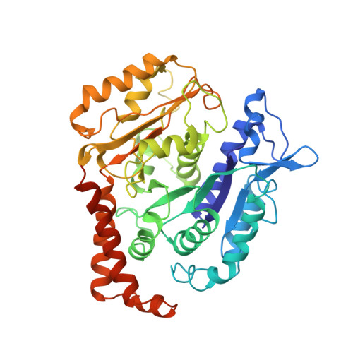
    6I2I

  • PubMed Abstract: 

    EML4 is a microtubule-associated protein that promotes microtubule stability. We investigated its regulation across the cell cycle and found that EML4 was distributed as punctate foci along the microtubule lattice in interphase but exhibited reduced association with spindle microtubules in mitosis. Microtubule sedimentation and cryo-electron microscopy with 3D reconstruction revealed that the basic N-terminal domain of EML4 mediated its binding to the acidic C-terminal tails of α- and β-tubulin on the microtubule surface. The mitotic kinases NEK6 and NEK7 phosphorylated the EML4 N-terminal domain at Ser 144 and Ser 146 in vitro, and depletion of these kinases in cells led to increased EML4 binding to microtubules in mitosis. An S144A-S146A double mutant not only bound inappropriately to mitotic microtubules but also increased their stability and interfered with chromosome congression. In addition, constitutive activation of NEK6 or NEK7 reduced the association of EML4 with interphase microtubules. Together, these data support a model in which NEK6- and NEK7-dependent phosphorylation promotes the dissociation of EML4 from microtubules in mitosis in a manner that is required for efficient chromosome congression.


  • Organizational Affiliation

    Department of Molecular and Cell Biology, University of Leicester, Lancaster Road, Leicester LE1 9HN, UK.


Macromolecules
Find similar proteins by:  (by identity cutoff)  |  3D Structure
Entity ID: 1
MoleculeChains Sequence LengthOrganismDetailsImage
Tubulin alpha-1B chain451Homo sapiensMutation(s): 0 
UniProt & NIH Common Fund Data Resources
Find proteins for P68363 (Homo sapiens)
Explore P68363 
Go to UniProtKB:  P68363
PHAROS:  P68363
GTEx:  ENSG00000123416 
Entity Groups  
Sequence Clusters30% Identity50% Identity70% Identity90% Identity95% Identity100% Identity
UniProt GroupP68363
Sequence Annotations
Expand
  • Reference Sequence
Find similar proteins by:  (by identity cutoff)  |  3D Structure
Entity ID: 2
MoleculeChains Sequence LengthOrganismDetailsImage
Tubulin beta chain444Homo sapiensMutation(s): 0 
UniProt & NIH Common Fund Data Resources
Find proteins for P07437 (Homo sapiens)
Explore P07437 
Go to UniProtKB:  P07437
PHAROS:  P07437
GTEx:  ENSG00000196230 
Entity Groups  
Sequence Clusters30% Identity50% Identity70% Identity90% Identity95% Identity100% Identity
UniProt GroupP07437
Sequence Annotations
Expand
  • Reference Sequence
Small Molecules
Binding Affinity Annotations 
IDSourceBinding Affinity
TA1 BindingDB:  6I2I EC50: 520 (nM) from 1 assay(s)
Experimental Data & Validation

Experimental Data

  • Method: ELECTRON MICROSCOPY
  • Resolution: 3.60 Å
  • Aggregation State: FILAMENT 
  • Reconstruction Method: SINGLE PARTICLE 
EM Software:
TaskSoftware PackageVersion
MODEL REFINEMENTPHENIX

Structure Validation

View Full Validation Report


Entry History & Funding Information

Deposition Data


Funding OrganizationLocationGrant Number
Wellcome TrustUnited Kingdom204801/Z/16/Z
United Kingdom16-0119
Medical Research Council (United Kingdom)United KingdomMR/R000352/1

Revision History  (Full details and data files)

  • Version 1.0: 2019-08-28
    Type: Initial release
  • Version 1.1: 2019-12-18
    Changes: Other