6LNG

Rapid crystallization of streptavidin using charged peptides


Experimental Data Snapshot

  • Method: X-RAY DIFFRACTION
  • Resolution: 1.80 Å
  • R-Value Free: 0.219 
  • R-Value Work: 0.181 
  • R-Value Observed: 0.183 

wwPDB Validation   3D Report Full Report


Ligand Structure Quality Assessment 


This is version 1.2 of the entry. See complete history


Literature

Genetically fused charged peptides induce rapid crystallization of proteins.

Minamihata, K.Tsukamoto, K.Adachi, M.Shimizu, R.Mishina, M.Kuroki, R.Nagamune, T.

(2020) Chem Commun (Camb) 56: 3891-3894

  • DOI: https://doi.org/10.1039/c9cc09529b
  • Primary Citation of Related Structures:  
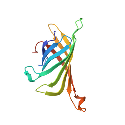
    6LNG

  • PubMed Abstract: 

    We utilized electrostatic interaction to induce rapid crystallization of streptavidin. Simply mixing streptavidins possessing either a positively or negatively charged peptide at their C-terminus generated diffraction-quality crystals in a few hours. We modified the streptavidin crystals with fluorescent molecules using biotin, demonstrating the concept of protein crystals as functional biomaterials.


  • Organizational Affiliation

    Department of Applied Chemistry, Graduate School of Engineering, Kyushu University, 744 Motooka, Fukuoka, 819-0395, Japan. kosukeminami@mail.cstm.kyushu-u.ac.jp.


Macromolecules
Find similar proteins by:  (by identity cutoff)  |  3D Structure
Entity ID: 1
MoleculeChains Sequence LengthOrganismDetailsImage
Streptavidin
A, B, C, D, E
A, B, C, D, E, F
125Streptomyces avidiniiMutation(s): 0 
UniProt
Find proteins for P22629 (Streptomyces avidinii)
Explore P22629 
Go to UniProtKB:  P22629
Entity Groups  
Sequence Clusters30% Identity50% Identity70% Identity90% Identity95% Identity100% Identity
UniProt GroupP22629
Sequence Annotations
Expand
  • Reference Sequence
Experimental Data & Validation

Experimental Data

  • Method: X-RAY DIFFRACTION
  • Resolution: 1.80 Å
  • R-Value Free: 0.219 
  • R-Value Work: 0.181 
  • R-Value Observed: 0.183 
  • Space Group: C 1 2 1
Unit Cell:
Length ( Å )Angle ( ˚ )
a = 81.2929992676α = 90
b = 80.8290023804β = 99.8460006714
c = 132.647994995γ = 90
Software Package:
Software NamePurpose
PHENIXrefinement
PDB_EXTRACTdata extraction
XDSdata reduction
SCALAdata scaling
PHASERphasing

Structure Validation

View Full Validation Report


Ligand Structure Quality Assessment 


Entry History 

Deposition Data

Revision History  (Full details and data files)

  • Version 1.0: 2020-03-18
    Type: Initial release
  • Version 1.1: 2020-04-15
    Changes: Database references
  • Version 1.2: 2023-11-22
    Changes: Data collection, Database references, Refinement description