6LR7

Crystal structure of GFPuv complexed with the nanobody LaG16 at 1.67 Angstron resolution


Experimental Data Snapshot

  • Method: X-RAY DIFFRACTION
  • Resolution: 1.67 Å
  • R-Value Free: 0.226 
  • R-Value Work: 0.185 
  • R-Value Observed: 0.187 

wwPDB Validation   3D Report Full Report


This is version 2.0 of the entry. See complete history


Literature

Structure-based engineering of anti-GFP nanobody tandems as ultra-high-affinity reagents for purification.

Zhang, Z.Wang, Y.Ding, Y.Hattori, M.

(2020) Sci Rep 10: 6239-6239

  • DOI: https://doi.org/10.1038/s41598-020-62606-7
  • Primary Citation of Related Structures:  
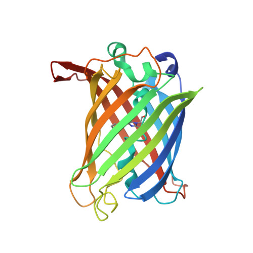
    6LR7

  • PubMed Abstract: 

    Green fluorescent proteins (GFPs) are widely used in biological research. Although GFP can be visualized easily, its precise manipulation through binding partners is still burdensome because of the limited availability of high-affinity binding partners and related structural information. Here, we report the crystal structure of GFPuv in complex with the anti-GFP nanobody LaG16 at 1.67 Å resolution, revealing the details of the binding between GFPuv and LaG16. The LaG16 binding site was on the opposite side of the GFP β-barrel from the binding site of the GFP-enhancer, another anti-GFP nanobody, indicating that the GFP-enhancer and LaG16 can bind to GFP together. Thus, we further designed 3 linkers of different lengths to fuse LaG16 and GFP-enhancer together, and the GFP binding of the three constructs was further tested by ITC. The construct with the (GGGGS) 4 linker had the highest affinity with a K D of 0.5 nM. The GFP-enhancer-(GGGGS) 4 -LaG16 chimeric nanobody was further covalently linked to NHS-activated agarose and then used in the purification of a GFP-tagged membrane protein, GFP-tagged zebrafish P2X4, resulting in higher yield than purification with the GFP-enhancer nanobody alone. This work provides a proof of concept for the design of ultra-high-affinity binders of target proteins through dimerized nanobody chimaeras, and this strategy may also be applied to link interesting target protein nanobodies without overlapping binding surfaces.


  • Organizational Affiliation

    State Key Laboratory of Genetic Engineering, Collaborative Innovation Center of Genetics and Development, Department of Physiology and Biophysics, School of Life Sciences, Fudan University, 2005 Songhu Road, Yangpu District, Shanghai, 200438, China.


Macromolecules
Find similar proteins by:  (by identity cutoff)  |  3D Structure
Entity ID: 1
MoleculeChains Sequence LengthOrganismDetailsImage
Green fluorescent protein239Aequorea victoriaMutation(s): 1 
Gene Names: GFP
UniProt
Find proteins for P42212 (Aequorea victoria)
Explore P42212 
Go to UniProtKB:  P42212
Entity Groups  
Sequence Clusters30% Identity50% Identity70% Identity90% Identity95% Identity100% Identity
UniProt GroupP42212
Sequence Annotations
Expand
  • Reference Sequence
Find similar proteins by:  (by identity cutoff)  |  3D Structure
Entity ID: 2
MoleculeChains Sequence LengthOrganismDetailsImage
Nanobody LaG16131Camelus bactrianusMutation(s): 0 
Entity Groups  
Sequence Clusters30% Identity50% Identity70% Identity90% Identity95% Identity100% Identity
Sequence Annotations
Expand
  • Reference Sequence
Small Molecules
Modified Residues  1 Unique
IDChains TypeFormula2D DiagramParent
CRO
Query on CRO
A
L-PEPTIDE LINKINGC15 H17 N3 O5THR, TYR, GLY
Experimental Data & Validation

Experimental Data

  • Method: X-RAY DIFFRACTION
  • Resolution: 1.67 Å
  • R-Value Free: 0.226 
  • R-Value Work: 0.185 
  • R-Value Observed: 0.187 
  • Space Group: P 1 21 1
Unit Cell:
Length ( Å )Angle ( ˚ )
a = 48.37α = 90
b = 41.78β = 91.89
c = 81.71γ = 90
Software Package:
Software NamePurpose
PHENIXrefinement
XSCALEdata scaling
PDB_EXTRACTdata extraction
XDSdata reduction
PHASERphasing

Structure Validation

View Full Validation Report


Entry History & Funding Information

Deposition Data


Funding OrganizationLocationGrant Number
Ministry of Science and Technology (MoST, China)China2016YFA0502800
National Natural Science Foundation of China (NSFC)China31850410466

Revision History  (Full details and data files)

  • Version 1.0: 2020-04-29
    Type: Initial release
  • Version 2.0: 2023-04-05
    Changes: Advisory, Atomic model, Author supporting evidence, Data collection, Database references, Derived calculations, Polymer sequence, Refinement description, Source and taxonomy, Structure summary