6MD4

Crystal Structure of Human PPARgamma Ligand Binding Domain in Complex with Rosiglitazone and Oleic acid


Experimental Data Snapshot

  • Method: X-RAY DIFFRACTION
  • Resolution: 2.24 Å
  • R-Value Free: 0.282 
  • R-Value Work: 0.240 
  • R-Value Observed: 0.242 

wwPDB Validation   3D Report Full Report

Currently 6MD4 does not have a validation slider image.


This is version 1.2 of the entry. See complete history


Literature

Cooperative cobinding of synthetic and natural ligands to the nuclear receptor PPAR gamma.

Shang, J.Brust, R.Mosure, S.A.Bass, J.Munoz-Tello, P.Lin, H.Hughes, T.S.Tang, M.Ge, Q.Kamekencka, T.M.Kojetin, D.J.

(2018) Elife 7

  • DOI: https://doi.org/10.7554/eLife.43320
  • Primary Citation of Related Structures:  
    5UGM, 6AUG, 6AVI, 6MCZ, 6MD0, 6MD1, 6MD2, 6MD4

  • PubMed Abstract: 

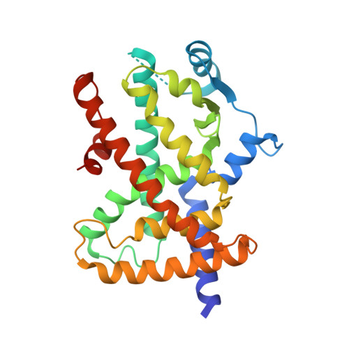
    Crystal structures of peroxisome proliferator-activated receptor gamma (PPARγ) have revealed overlapping binding modes for synthetic and natural/endogenous ligands, indicating competition for the orthosteric pocket. Here we show that cobinding of a synthetic ligand to the orthosteric pocket can push natural and endogenous PPARγ ligands (fatty acids) out of the orthosteric pocket towards an alternate ligand-binding site near the functionally important omega (Ω)-loop. X-ray crystallography, NMR spectroscopy, all-atom molecular dynamics simulations, and mutagenesis coupled to quantitative biochemical functional and cellular assays reveal that synthetic ligand and fatty acid cobinding can form a 'ligand link' to the Ω-loop and synergistically affect the structure and function of PPARγ. These findings contribute to a growing body of evidence indicating ligand binding to nuclear receptors can be more complex than the classical one-for-one orthosteric exchange of a natural or endogenous ligand with a synthetic ligand.


  • Organizational Affiliation

    Department of Integrative Structural and Computational Biology, The Scripps Research Institute, Jupiter, United States.


Macromolecules
Find similar proteins by:  (by identity cutoff)  |  3D Structure
Entity ID: 1
MoleculeChains Sequence LengthOrganismDetailsImage
Peroxisome proliferator-activated receptor gamma
A, B
275Homo sapiensMutation(s): 0 
Gene Names: PPARGNR1C3
UniProt & NIH Common Fund Data Resources
Find proteins for P37231 (Homo sapiens)
Explore P37231 
Go to UniProtKB:  P37231
PHAROS:  P37231
GTEx:  ENSG00000132170 
Entity Groups  
Sequence Clusters30% Identity50% Identity70% Identity90% Identity95% Identity100% Identity
UniProt GroupP37231
Sequence Annotations
Expand
  • Reference Sequence
Small Molecules
Ligands 2 Unique
IDChains Name / Formula / InChI Key2D Diagram3D Interactions
BRL
Query on BRL

Download Ideal Coordinates CCD File 
C [auth A]2,4-THIAZOLIDIINEDIONE, 5-[[4-[2-(METHYL-2-PYRIDINYLAMINO)ETHOXY]PHENYL]METHYL]-(9CL)
C18 H19 N3 O3 S
YASAKCUCGLMORW-HNNXBMFYSA-N
OLA
Query on OLA

Download Ideal Coordinates CCD File 
D [auth A]OLEIC ACID
C18 H34 O2
ZQPPMHVWECSIRJ-KTKRTIGZSA-N
Binding Affinity Annotations 
IDSourceBinding Affinity
BRL BindingDB:  6MD4 Ki: min: 8, max: 59 (nM) from 5 assay(s)
Kd: min: 0.14, max: 48 (nM) from 3 assay(s)
IC50: min: 1, max: 298 (nM) from 10 assay(s)
EC50: min: 1, max: 1600 (nM) from 35 assay(s)
OLA BindingDB:  6MD4 IC50: 4100 (nM) from 1 assay(s)
Experimental Data & Validation

Experimental Data

  • Method: X-RAY DIFFRACTION
  • Resolution: 2.24 Å
  • R-Value Free: 0.282 
  • R-Value Work: 0.240 
  • R-Value Observed: 0.242 
  • Space Group: C 1 2 1
Unit Cell:
Length ( Å )Angle ( ˚ )
a = 92.829α = 90
b = 61.834β = 102.34
c = 118.717γ = 90
Software Package:
Software NamePurpose
PHENIXrefinement
Aimlessdata scaling
PDB_EXTRACTdata extraction
MOSFLMdata reduction
PHASERphasing

Structure Validation

View Full Validation Report

Currently 6MD4 does not have a validation slider image.


Entry History & Funding Information

Deposition Data


Funding OrganizationLocationGrant Number
National Institutes of Health/National Institute of Diabetes and Digestive and Kidney Disease (NIH/NIDDK)United StatesR01DK101871

Revision History  (Full details and data files)

  • Version 1.0: 2019-01-09
    Type: Initial release
  • Version 1.1: 2019-12-25
    Changes: Author supporting evidence
  • Version 1.2: 2023-10-11
    Changes: Data collection, Database references, Refinement description