6QL1

Crystal structure of chimeric carbonic anhydrase VI with 4-[(4,6-dimethylpyrimidin-2-yl)thio]-2,3,5,6-tetrafluorobenzenesulfonamide


Experimental Data Snapshot

  • Method: X-RAY DIFFRACTION
  • Resolution: 1.42 Å
  • R-Value Free: 0.186 
  • R-Value Work: 0.158 
  • R-Value Observed: 0.161 

wwPDB Validation   3D Report Full Report


Ligand Structure Quality Assessment 


This is version 1.1 of the entry. See complete history


Literature

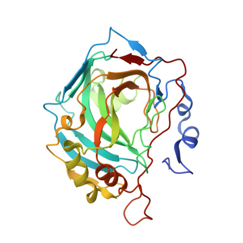
Engineered Carbonic Anhydrase VI-Mimic Enzyme Switched the Structure and Affinities of Inhibitors.

Kazokaite, J.Kairys, V.Smirnoviene, J.Smirnov, A.Manakova, E.Tolvanen, M.Parkkila, S.Matulis, D.

(2019) Sci Rep 9: 12710-12710

  • DOI: https://doi.org/10.1038/s41598-019-49094-0
  • Primary Citation of Related Structures:  
    6QL1, 6QL2, 6QL3

  • PubMed Abstract: 

    Secretory human carbonic anhydrase VI (CA VI) has emerged as a potential drug target due to its role in pathological states, such as excess acidity-caused dental caries and injuries of gastric epithelium. Currently, there are no available CA VI-selective inhibitors or crystallographic structures of inhibitors bound to CA VI. The present study focuses on the site-directed CA II mutant mimicking the active site of CA VI for inhibitor screening. The interactions between CA VI-mimic and a series of benzenesulfonamides were evaluated by fluorescent thermal shift assay, stopped-flow CO 2 hydration assay, isothermal titration calorimetry, and X-ray crystallography. Kinetic parameters showed that A65T, N67Q, F130Y, V134Q, L203T mutations did not influence catalytic properties of CA II, but inhibitor affinities resembled CA VI, exhibiting up to 0.16 nM intrinsic affinity for CA VI-mimic. Structurally, binding site of CA VI-mimic was found to be similar to CA VI. The ligand interactions with mutated side chains observed in three crystallographic structures allowed to rationalize observed variation of binding modes and experimental binding affinities to CA VI. This integrative set of kinetic, thermodynamic, and structural data revealed CA VI-mimic as a useful model to design CA VI-specific inhibitors which could be beneficial for novel therapeutic applications.


  • Organizational Affiliation

    Department of Biothermodynamics and Drug Design, Institute of Biotechnology, Vilnius University, Saulėtekio 7, Vilnius, LT-10257, Lithuania. kazokaite@ibt.lt.


Macromolecules
Find similar proteins by:  (by identity cutoff)  |  3D Structure
Entity ID: 1
MoleculeChains Sequence LengthOrganismDetailsImage
Carbonic anhydrase 2260Homo sapiensMutation(s): 5 
Gene Names: CA2
EC: 4.2.1.1
UniProt & NIH Common Fund Data Resources
Find proteins for P00918 (Homo sapiens)
Explore P00918 
Go to UniProtKB:  P00918
PHAROS:  P00918
GTEx:  ENSG00000104267 
Entity Groups  
Sequence Clusters30% Identity50% Identity70% Identity90% Identity95% Identity100% Identity
UniProt GroupP00918
Sequence Annotations
Expand
  • Reference Sequence
Small Molecules
Ligands 6 Unique
IDChains Name / Formula / InChI Key2D Diagram3D Interactions
V50
Query on V50

Download Ideal Coordinates CCD File 
F [auth A],
G [auth A]
4-[(4,6-dimethylpyrimidin-2-yl)thio]-2,3,5,6-tetrafluorobenzenesulfonamide
C12 H9 F4 N3 O2 S2
PKQIZTGHFBCYEN-UHFFFAOYSA-N
BCN
Query on BCN

Download Ideal Coordinates CCD File 
E [auth A]BICINE
C6 H13 N O4
FSVCELGFZIQNCK-UHFFFAOYSA-N
DMS
Query on DMS

Download Ideal Coordinates CCD File 
C [auth A]DIMETHYL SULFOXIDE
C2 H6 O S
IAZDPXIOMUYVGZ-UHFFFAOYSA-N
ZN
Query on ZN

Download Ideal Coordinates CCD File 
B [auth A]ZINC ION
Zn
PTFCDOFLOPIGGS-UHFFFAOYSA-N
EDO
Query on EDO

Download Ideal Coordinates CCD File 
D [auth A]1,2-ETHANEDIOL
C2 H6 O2
LYCAIKOWRPUZTN-UHFFFAOYSA-N
NA
Query on NA

Download Ideal Coordinates CCD File 
H [auth A]SODIUM ION
Na
FKNQFGJONOIPTF-UHFFFAOYSA-N
Binding Affinity Annotations 
IDSourceBinding Affinity
V50 BindingDB:  6QL1 Kd: 10 (nM) from 1 assay(s)
Binding MOAD:  6QL1 Kd: 15 (nM) from 1 assay(s)
Experimental Data & Validation

Experimental Data

  • Method: X-RAY DIFFRACTION
  • Resolution: 1.42 Å
  • R-Value Free: 0.186 
  • R-Value Work: 0.158 
  • R-Value Observed: 0.161 
  • Space Group: P 1 21 1
Unit Cell:
Length ( Å )Angle ( ˚ )
a = 42.299α = 90
b = 41.401β = 104.31
c = 71.242γ = 90
Software Package:
Software NamePurpose
REFMACrefinement
PDB_EXTRACTdata extraction
XDSdata reduction
SCALAdata scaling
MOLREPphasing
Cootmodel building

Structure Validation

View Full Validation Report


Ligand Structure Quality Assessment 


Entry History 

Deposition Data

Revision History  (Full details and data files)

  • Version 1.0: 2019-09-25
    Type: Initial release
  • Version 1.1: 2024-01-24
    Changes: Advisory, Data collection, Database references, Derived calculations, Refinement description