6QVJ

HsCKK (human CAMSAP1) decorated 14pf taxol-GDP microtubule


Experimental Data Snapshot

  • Method: ELECTRON MICROSCOPY
  • Resolution: 3.80 Å
  • Aggregation State: FILAMENT 
  • Reconstruction Method: SINGLE PARTICLE 

wwPDB Validation   3D Report Full Report


This is version 1.4 of the entry. See complete history


Literature

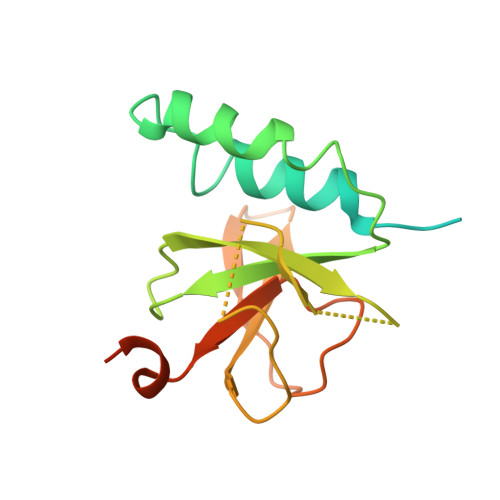
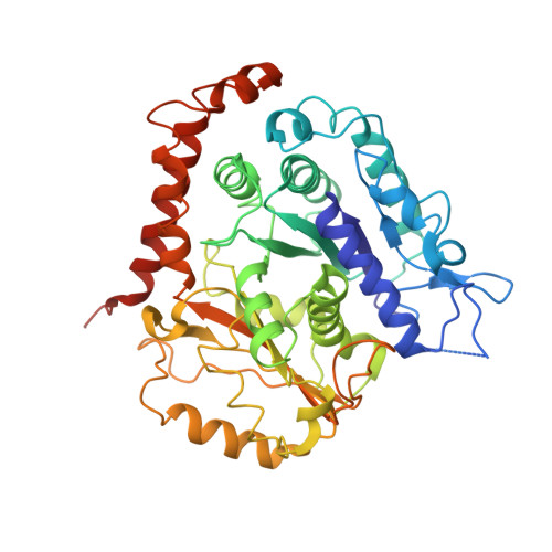
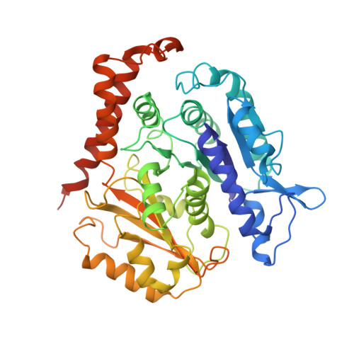
Structural determinants of microtubule minus end preference in CAMSAP CKK domains.

Atherton, J.Luo, Y.Xiang, S.Yang, C.Rai, A.Jiang, K.Stangier, M.Vemu, A.Cook, A.D.Wang, S.Roll-Mecak, A.Steinmetz, M.O.Akhmanova, A.Baldus, M.Moores, C.A.

(2019) Nat Commun 10: 5236-5236

  • DOI: https://doi.org/10.1038/s41467-019-13247-6
  • Primary Citation of Related Structures:  
    6QUS, 6QUY, 6QVE, 6QVJ

  • PubMed Abstract: 

    CAMSAP/Patronins regulate microtubule minus-end dynamics. Their end specificity is mediated by their CKK domains, which we proposed recognise specific tubulin conformations found at minus ends. To critically test this idea, we compared the human CAMSAP1 CKK domain (HsCKK) with a CKK domain from Naegleria gruberi (NgCKK), which lacks minus-end specificity. Here we report near-atomic cryo-electron microscopy structures of HsCKK- and NgCKK-microtubule complexes, which show that these CKK domains share the same protein fold, bind at the intradimer interprotofilament tubulin junction, but exhibit different footprints on microtubules. NMR experiments show that both HsCKK and NgCKK are remarkably rigid. However, whereas NgCKK binding does not alter the microtubule architecture, HsCKK remodels its microtubule interaction site and changes the underlying polymer structure because the tubulin lattice conformation is not optimal for its binding. Thus, in contrast to many MAPs, the HsCKK domain can differentiate subtly specific tubulin conformations to enable microtubule minus-end recognition.


  • Organizational Affiliation

    Institute of Structural and Molecular Biology, Birkbeck, University of London, Malet Street, London, UK. j.atherton@mail.cryst.bbk.ac.uk.


Macromolecules
Find similar proteins by:  (by identity cutoff)  |  3D Structure
Entity ID: 1
MoleculeChains Sequence LengthOrganismDetailsImage
Calmodulin-regulated spectrin-associated protein 1A [auth I]174Homo sapiensMutation(s): 0 
Gene Names: CAMSAP1
UniProt & NIH Common Fund Data Resources
Find proteins for Q5T5Y3 (Homo sapiens)
Explore Q5T5Y3 
Go to UniProtKB:  Q5T5Y3
PHAROS:  Q5T5Y3
GTEx:  ENSG00000130559 
Entity Groups  
Sequence Clusters30% Identity50% Identity70% Identity90% Identity95% Identity100% Identity
UniProt GroupQ5T5Y3
Sequence Annotations
Expand
  • Reference Sequence
Find similar proteins by:  (by identity cutoff)  |  3D Structure
Entity ID: 2
MoleculeChains Sequence LengthOrganismDetailsImage
Tubulin alpha-1B chainB [auth O],
C [auth X]
451Homo sapiensMutation(s): 0 
UniProt & NIH Common Fund Data Resources
Find proteins for P68363 (Homo sapiens)
Explore P68363 
Go to UniProtKB:  P68363
PHAROS:  P68363
GTEx:  ENSG00000123416 
Entity Groups  
Sequence Clusters30% Identity50% Identity70% Identity90% Identity95% Identity100% Identity
UniProt GroupP68363
Sequence Annotations
Expand
  • Reference Sequence
Find similar proteins by:  (by identity cutoff)  |  3D Structure
Entity ID: 3
MoleculeChains Sequence LengthOrganismDetailsImage
Tubulin beta chainD [auth S],
E [auth U]
444Homo sapiensMutation(s): 0 
UniProt & NIH Common Fund Data Resources
Find proteins for P07437 (Homo sapiens)
Explore P07437 
Go to UniProtKB:  P07437
PHAROS:  P07437
GTEx:  ENSG00000196230 
Entity Groups  
Sequence Clusters30% Identity50% Identity70% Identity90% Identity95% Identity100% Identity
UniProt GroupP07437
Sequence Annotations
Expand
  • Reference Sequence
Small Molecules
Ligands 4 Unique
IDChains Name / Formula / InChI Key2D Diagram3D Interactions
TA1
Query on TA1

Download Ideal Coordinates CCD File 
K [auth S],
M [auth U]
TAXOL
C47 H51 N O14
RCINICONZNJXQF-MZXODVADSA-N
GTP
Query on GTP

Download Ideal Coordinates CCD File 
F [auth O],
H [auth X]
GUANOSINE-5'-TRIPHOSPHATE
C10 H16 N5 O14 P3
XKMLYUALXHKNFT-UUOKFMHZSA-N
GDP
Query on GDP

Download Ideal Coordinates CCD File 
J [auth S],
L [auth U]
GUANOSINE-5'-DIPHOSPHATE
C10 H15 N5 O11 P2
QGWNDRXFNXRZMB-UUOKFMHZSA-N
MG
Query on MG

Download Ideal Coordinates CCD File 
G [auth O],
I [auth X]
MAGNESIUM ION
Mg
JLVVSXFLKOJNIY-UHFFFAOYSA-N
Binding Affinity Annotations 
IDSourceBinding Affinity
TA1 BindingDB:  6QVJ EC50: 520 (nM) from 1 assay(s)
Experimental Data & Validation

Experimental Data

  • Method: ELECTRON MICROSCOPY
  • Resolution: 3.80 Å
  • Aggregation State: FILAMENT 
  • Reconstruction Method: SINGLE PARTICLE 
EM Software:
TaskSoftware PackageVersion
MODEL REFINEMENTPHENIX

Structure Validation

View Full Validation Report


Entry History & Funding Information

Deposition Data


Funding OrganizationLocationGrant Number
Medical Research Council (United Kingdom)United KingdomMR/R000352/1
Netherlands722.016.002
Netherlands175.010.2009.002
Netherlands718.015.001
Netherlands184.032.207
Swiss National Science FoundationSwitzerland31003A_166608

Revision History  (Full details and data files)

  • Version 1.0: 2019-11-27
    Type: Initial release
  • Version 1.1: 2019-12-04
    Changes: Database references
  • Version 1.2: 2019-12-18
    Changes: Other
  • Version 1.3: 2020-08-26
    Changes: Database references, Derived calculations
  • Version 1.4: 2020-09-09
    Changes: Database references