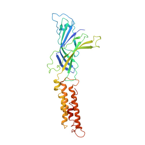
Cryo-EM structures of a lipid-sensitive pentameric ligand-gated ion channel embedded in a phosphatidylcholine-only bilayer.
Kumar, P., Wang, Y., Zhang, Z., Zhao, Z., Cymes, G.D., Tajkhorshid, E., Grosman, C.(2020) Proc Natl Acad Sci U S A 117: 1788-1798
- PubMed: 31911476
- DOI: https://doi.org/10.1073/pnas.1906823117
- Primary Citation of Related Structures:
6V03, 6V0B - PubMed Abstract:
The lipid dependence of the nicotinic acetylcholine receptor from the Torpedo electric organ has long been recognized, and one of the most consistent experimental observations is that, when reconstituted in membranes formed by zwitterionic phospholipids alone, exposure to agonist fails to elicit ion-flux activity. More recently, it has been suggested that the bacterial homolog ELIC ( Erwinia chrysanthemi ligand-gated ion channel) has a similar lipid sensitivity. As a first step toward the elucidation of the structural basis of this phenomenon, we solved the structures of ELIC embedded in palmitoyl-oleoyl-phosphatidylcholine- (POPC-) only nanodiscs in both the unliganded (4.1-Å resolution) and agonist-bound (3.3 Å) states using single-particle cryoelectron microscopy. Comparison of the two structural models revealed that the largest differences occur at the level of loop C-at the agonist-binding sites-and the loops at the interface between the extracellular and transmembrane domains (ECD and TMD, respectively). On the other hand, the transmembrane pore is occluded in a remarkably similar manner in both structures. A straightforward interpretation of these findings is that POPC-only membranes frustrate the ECD-TMD coupling in such a way that the "conformational wave" of liganded-receptor gating takes place in the ECD and the interfacial M2-M3 linker but fails to penetrate the membrane and propagate into the TMD. Furthermore, analysis of the structural models and molecular simulations suggested that the higher affinity for agonists characteristic of the open- and desensitized-channel conformations results, at least in part, from the tighter confinement of the ligand to its binding site; this limits the ligand's fluctuations, and thus delays its escape into bulk solvent.
Organizational Affiliation:
Department of Molecular and Integrative Physiology, University of Illinois at Urbana-Champaign, Urbana, IL 61801.