6W4E

NMR-driven structure of KRAS4B-GTP homodimer on a lipid bilayer nanodisc


Experimental Data Snapshot

  • Method: SOLUTION NMR
  • Conformers Calculated: 3000 
  • Conformers Submitted: 20 
  • Selection Criteria: structures with the lowest energy 

wwPDB Validation   3D Report Full Report


This is version 1.1 of the entry. See complete history


Literature

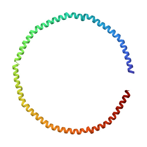
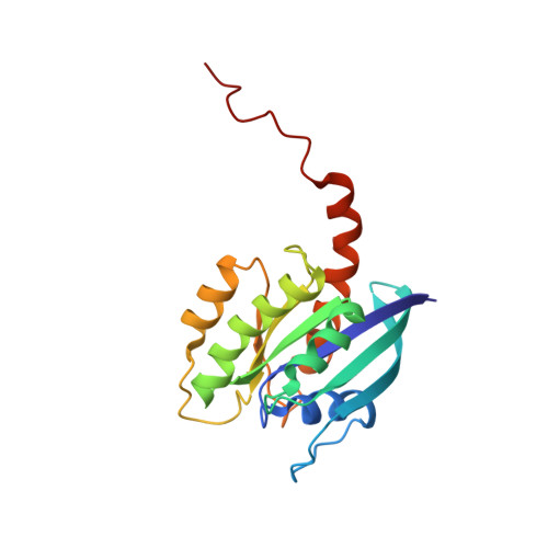
Two Distinct Structures of Membrane-Associated Homodimers of GTP- and GDP-Bound KRAS4B Revealed by Paramagnetic Relaxation Enhancement.

Lee, K.Y.Fang, Z.Enomoto, M.Gasmi-Seabrook, G.Zheng, L.Koide, S.Ikura, M.Marshall, C.B.

(2020) Angew Chem Int Ed Engl 59: 11037-11045

  • DOI: https://doi.org/10.1002/anie.202001758
  • Primary Citation of Related Structures:  
    6W4E, 6W4F

  • PubMed Abstract: 

    KRAS homo-dimerization has been implicated in the activation of RAF kinases, however, the mechanism and structural basis remain elusive. We developed a system to study KRAS dimerization on nanodiscs using paramagnetic relaxation enhancement (PRE) NMR spectroscopy, and determined distinct structures of membrane-anchored KRAS dimers in the active GTP- and inactive GDP-loaded states. Both dimerize through an α4-α5 interface, but the relative orientation of the protomers and their contacts differ substantially. Dimerization of KRAS-GTP, stabilized by electrostatic interactions between R135 and E168, favors an orientation on the membrane that promotes accessibility of the effector-binding site. Remarkably, "cross"-dimerization between GTP- and GDP-bound KRAS molecules is unfavorable. These models provide a platform to elucidate the structural basis of RAF activation by RAS and to develop inhibitors that can disrupt the KRAS dimerization. The methodology is applicable to many other farnesylated small GTPases.


  • Organizational Affiliation

    Princess Margaret Cancer Centre, University Health Network, Toronto, Ontario, M5G 1L7, Canada.


Macromolecules
Find similar proteins by:  (by identity cutoff)  |  3D Structure
Entity ID: 1
MoleculeChains Sequence LengthOrganismDetailsImage
Apolipoprotein A-IA,
B [auth D]
200Homo sapiensMutation(s): 0 
Gene Names: APOA1
UniProt & NIH Common Fund Data Resources
Find proteins for P02647 (Homo sapiens)
Explore P02647 
Go to UniProtKB:  P02647
PHAROS:  P02647
GTEx:  ENSG00000118137 
Entity Groups  
Sequence Clusters30% Identity50% Identity70% Identity90% Identity95% Identity100% Identity
UniProt GroupP02647
Sequence Annotations
Expand
  • Reference Sequence
Find similar proteins by:  (by identity cutoff)  |  3D Structure
Entity ID: 2
MoleculeChains Sequence LengthOrganismDetailsImage
GTPase KRasC [auth B],
D [auth C]
185Homo sapiensMutation(s): 0 
Gene Names: KRASKRAS2RASK2
UniProt & NIH Common Fund Data Resources
Find proteins for P01116 (Homo sapiens)
Explore P01116 
Go to UniProtKB:  P01116
PHAROS:  P01116
GTEx:  ENSG00000133703 
Entity Groups  
Sequence Clusters30% Identity50% Identity70% Identity90% Identity95% Identity100% Identity
UniProt GroupP01116
Sequence Annotations
Expand
  • Reference Sequence
Small Molecules
Ligands 4 Unique
IDChains Name / Formula / InChI Key2D Diagram3D Interactions
17F
Query on 17F

Download Ideal Coordinates CCD File 
BB [auth D]
CB [auth D]
DB [auth D]
DC [auth C]
EB [auth D]
BB [auth D],
CB [auth D],
DB [auth D],
DC [auth C],
EB [auth D],
EC [auth C],
FB [auth D],
FC [auth C],
IC [auth C],
JC [auth C],
OB [auth B],
PB [auth B],
QB [auth B],
S [auth A],
T [auth A],
Z [auth A]
O-[(S)-({(2R)-2,3-bis[(9Z)-octadec-9-enoyloxy]propyl}oxy)(hydroxy)phosphoryl]-L-serine
C42 H78 N O10 P
WTBFLCSPLLEDEM-JIDRGYQWSA-N
PCW
Query on PCW

Download Ideal Coordinates CCD File 
AA [auth D]
AB [auth D]
AC [auth C]
BA [auth D]
BC [auth C]
AA [auth D],
AB [auth D],
AC [auth C],
BA [auth D],
BC [auth C],
CA [auth D],
CC [auth C],
DA [auth D],
E [auth A],
EA [auth D],
F [auth A],
FA [auth D],
G [auth A],
GA [auth D],
GC [auth C],
H [auth A],
HA [auth D],
HC [auth C],
I [auth A],
IA [auth D],
IB [auth B],
J [auth A],
JA [auth D],
JB [auth B],
K [auth A],
KA [auth D],
KB [auth B],
L [auth A],
LA [auth D],
LB [auth B],
M [auth A],
MA [auth D],
MB [auth B],
N [auth A],
NA [auth D],
NB [auth B],
O [auth A],
OA [auth D],
P [auth A],
PA [auth D],
Q [auth A],
QA [auth D],
R [auth A],
RA [auth D],
SA [auth D],
TA [auth D],
TB [auth C],
U [auth A],
UA [auth D],
UB [auth C],
V [auth A],
VA [auth D],
VB [auth C],
W [auth A],
WA [auth D],
WB [auth C],
X [auth A],
XA [auth D],
XB [auth C],
Y [auth A],
YA [auth D],
YB [auth C],
ZA [auth D],
ZB [auth C]
1,2-DIOLEOYL-SN-GLYCERO-3-PHOSPHOCHOLINE
C44 H85 N O8 P
SNKAWJBJQDLSFF-NVKMUCNASA-O
GSP
Query on GSP

Download Ideal Coordinates CCD File 
GB [auth B],
RB [auth C]
5'-GUANOSINE-DIPHOSPHATE-MONOTHIOPHOSPHATE
C10 H16 N5 O13 P3 S
XOFLBQFBSOEHOG-UUOKFMHZSA-N
MG
Query on MG

Download Ideal Coordinates CCD File 
HB [auth B],
SB [auth C]
MAGNESIUM ION
Mg
JLVVSXFLKOJNIY-UHFFFAOYSA-N
Experimental Data & Validation

Experimental Data

  • Method: SOLUTION NMR
  • Conformers Calculated: 3000 
  • Conformers Submitted: 20 
  • Selection Criteria: structures with the lowest energy 

Structure Validation

View Full Validation Report


Entry History & Funding Information

Deposition Data


Funding OrganizationLocationGrant Number
Canadian Institutes of Health Research (CIHR)Canada--

Revision History  (Full details and data files)

  • Version 1.0: 2020-04-15
    Type: Initial release
  • Version 1.1: 2020-07-08
    Changes: Database references