Protein structural changes on a CubeSat under rocket acceleration profile.
Luna, A., Meisel, J., Hsu, K., Russi, S., Fernandez, D.(2020) NPJ Microgravity 6: 12-12
- PubMed: 32352028
- DOI: https://doi.org/10.1038/s41526-020-0102-3
- Primary Citation of Related Structures:
6W7P, 6W8E - PubMed Abstract:
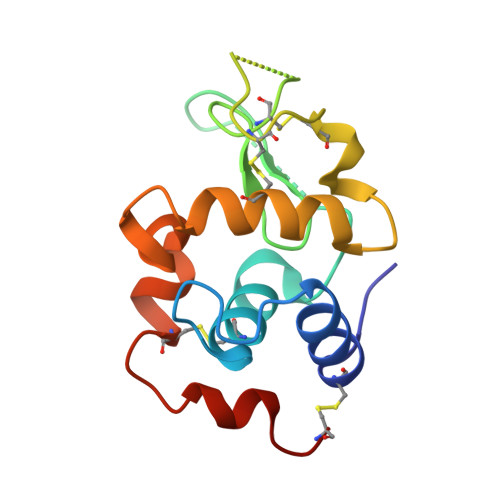
Catalyzing life-sustaining reactions, proteins are composed by 20 different amino acids that fold into a compact yet flexible three-dimensional architecture, which dictates what their function(s) might be. Determining the spatial arrangement of the atoms, the protein's 3D structure, enables key advances in fundamental and applied research. Protein crystallization is a powerful technique to achieve this. Unlike Earth's crystallization experiments, biomolecular crystallization in space in the absence of gravitational force is actively sought to improve crystal growth techniques. However, the effects of changing gravitational vectors on a protein solution reaching supersaturation remain largely unknown. Here, we have developed a low-cost crystallization cell within a CubeSat payload module to exploit the unique experimental conditions set aboard a sounding rocket. We designed a biaxial gimbal to house the crystallization experiments and take measurements on the protein solution in-flight with a spectrophotometry system. After flight, we used X-ray diffraction analysis to determine that flown protein has a structural rearrangement marked by loss of the protein's water and sodium in a manner that differs from crystals grown on the ground. We finally show that our gimbal payload module design is a portable experimental setup to take laboratory research investigations into exploratory space flights.
Organizational Affiliation:
1Mechanical Engineering Department, School of Engineering, Stanford University, Stanford, CA 94305 USA.