6Y4T

Crystal structure of p38 in complex with SR63.


Experimental Data Snapshot

  • Method: X-RAY DIFFRACTION
  • Resolution: 1.98 Å
  • R-Value Free: 0.251 
  • R-Value Work: 0.198 
  • R-Value Observed: 0.201 

wwPDB Validation   3D Report Full Report


Ligand Structure Quality Assessment 


This is version 1.3 of the entry. See complete history


Literature

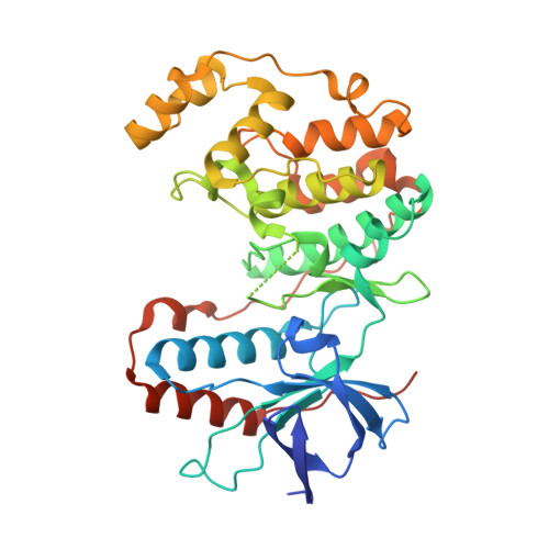
Selective targeting of the alpha C and DFG-out pocket in p38 MAPK.

Rohm, S.Schroder, M.Dwyer, J.E.Widdowson, C.S.Chaikuad, A.Berger, B.T.Joerger, A.C.Kramer, A.Harbig, J.Dauch, D.Kudolo, M.Laufer, S.Bagley, M.C.Knapp, S.

(2020) Eur J Med Chem 208: 112721-112721

  • DOI: https://doi.org/10.1016/j.ejmech.2020.112721
  • Primary Citation of Related Structures:  
    6Y4T, 6Y4U, 6Y4V, 6Y4W, 6Y4X, 6Y6V, 6YJC, 6YK7, 6ZWP, 6ZWR

  • PubMed Abstract: 

    The p38 MAPK cascade is a key signaling pathway linked to a multitude of physiological functions and of central importance in inflammatory and autoimmune diseases. Although studied extensively, little is known about how conformation-specific inhibitors alter signaling outcomes. Here, we have explored the highly dynamic back pocket of p38 MAPK with allosteric urea fragments. However, screening against known off-targets showed that these fragments maintained the selectivity issues of their parent compound BIRB-796, while combination with the hinge-binding motif of VPC-00628 greatly enhanced inhibitor selectivity. Further efforts focused therefore on the exploration of the αC-out pocket of p38 MAPK, yielding compound 137 as a highly selective type-II inhibitor. Even though 137 is structurally related to a recent p38 type-II chemical probe, SR-318, the data presented here provide valuable insights into back-pocket interactions that are not addressed in SR-318 and it provides an alternative chemical tool with good cellular activity targeting also the p38 back pocket.


  • Organizational Affiliation

    Johann Wolfgang Goethe University, Institute of Pharmaceutical Chemistry, Max-von-Laue-Str. 9, 60438, Frankfurt Am Main, Germany; Buchmann Institute for Molecular Life Sciences and Structural Genomics Consortium (SGC), Max-von-Laue-Str. 15, 60438, Frankfurt Am Main, Germany.


Macromolecules
Find similar proteins by:  (by identity cutoff)  |  3D Structure
Entity ID: 1
MoleculeChains Sequence LengthOrganismDetailsImage
Mitogen-activated protein kinase 14361Mus musculusMutation(s): 0 
Gene Names: Mapk14Crk1Csbp1Csbp2
EC: 2.7.11.24
UniProt
Find proteins for P47811 (Mus musculus)
Explore P47811 
Go to UniProtKB:  P47811
Entity Groups  
Sequence Clusters30% Identity50% Identity70% Identity90% Identity95% Identity100% Identity
UniProt GroupP47811
Sequence Annotations
Expand
  • Reference Sequence
Small Molecules
Ligands 2 Unique
IDChains Name / Formula / InChI Key2D Diagram3D Interactions
O8W (Subject of Investigation/LOI)
Query on O8W

Download Ideal Coordinates CCD File 
B [auth A]5-azanyl-~{N}-[[4-[[(2~{S})-1-[[(2~{S})-butan-2-yl]amino]-4-cyclohexyl-1-oxidanylidene-butan-2-yl]carbamoyl]phenyl]methyl]-1-phenyl-pyrazole-4-carboxamide
C32 H42 N6 O3
XHAOZPSJXRBUJD-DWACAAAGSA-N
EDO
Query on EDO

Download Ideal Coordinates CCD File 
C [auth A]
D [auth A]
E [auth A]
F [auth A]
G [auth A]
C [auth A],
D [auth A],
E [auth A],
F [auth A],
G [auth A],
H [auth A],
I [auth A]
1,2-ETHANEDIOL
C2 H6 O2
LYCAIKOWRPUZTN-UHFFFAOYSA-N
Experimental Data & Validation

Experimental Data

  • Method: X-RAY DIFFRACTION
  • Resolution: 1.98 Å
  • R-Value Free: 0.251 
  • R-Value Work: 0.198 
  • R-Value Observed: 0.201 
  • Space Group: P 21 21 21
Unit Cell:
Length ( Å )Angle ( ˚ )
a = 66.079α = 90
b = 75.03β = 90
c = 77.89γ = 90
Software Package:
Software NamePurpose
SCALAdata scaling
REFMACrefinement
PDB_EXTRACTdata extraction
iMOSFLMdata reduction
PHASERphasing

Structure Validation

View Full Validation Report


Ligand Structure Quality Assessment 


Entry History 

Deposition Data

Revision History  (Full details and data files)

  • Version 1.0: 2020-03-04
    Type: Initial release
  • Version 1.1: 2020-04-15
    Changes: Structure summary
  • Version 1.2: 2021-01-13
    Changes: Database references
  • Version 1.3: 2024-01-24
    Changes: Data collection, Database references, Refinement description