7RSA

STRUCTURE OF PHOSPHATE-FREE RIBONUCLEASE A REFINED AT 1.26 ANGSTROMS


Experimental Data Snapshot

  • Method: X-RAY DIFFRACTION
  • Resolution: 1.26 Å
  • R-Value Observed: 0.150 

wwPDB Validation   3D Report Full Report


This is version 2.0 of the entry. See complete history


Literature

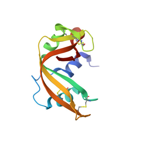
Structure of phosphate-free ribonuclease A refined at 1.26 A.

Wlodawer, A.Svensson, L.A.Sjolin, L.Gilliland, G.L.

(1988) Biochemistry 27: 2705-2717

  • DOI: https://doi.org/10.1021/bi00408a010
  • Primary Citation of Related Structures:  
    7RSA

  • PubMed Abstract: 

    The structure of phosphate-free bovine ribonuclease A has been refined at 1.26-A resolution by a restrained least-squares procedure to a final R factor of 0.15. X-ray diffraction data were collected with an electronic position-sensitive detector. The final model consists of all atoms in the polypeptide chain including hydrogens, 188 water sites with full or partial occupancy, and a single molecule of 2-methyl-2-propanol. Thirteen side chains were modeled with two alternate conformations. Major changes to the active site include the addition of two waters in the phosphate-binding pocket, disordering of Gln-11, and tilting of the imidazole ring of His-119. The structure of the protein and of the associated solvent was extensively compared with three other high-resolution, refined structures of this enzyme.


  • Organizational Affiliation

    Center for Chemical Physics, National Bureau of Standards, Gaithersburg, Maryland 20899.


Macromolecules
Find similar proteins by:  (by identity cutoff)  |  3D Structure
Entity ID: 1
MoleculeChains Sequence LengthOrganismDetailsImage
RIBONUCLEASE A124Bos taurusMutation(s): 0 
EC: 3.1.27.5
UniProt
Find proteins for P61823 (Bos taurus)
Explore P61823 
Go to UniProtKB:  P61823
Entity Groups  
Sequence Clusters30% Identity50% Identity70% Identity90% Identity95% Identity100% Identity
UniProt GroupP61823
Sequence Annotations
Expand
  • Reference Sequence
Small Molecules
Ligands 1 Unique
IDChains Name / Formula / InChI Key2D Diagram3D Interactions
TBU
Query on TBU

Download Ideal Coordinates CCD File 
B [auth A]TERTIARY-BUTYL ALCOHOL
C4 H10 O
DKGAVHZHDRPRBM-UHFFFAOYSA-N
Experimental Data & Validation

Experimental Data

  • Method: X-RAY DIFFRACTION
  • Resolution: 1.26 Å
  • R-Value Observed: 0.150 
  • Space Group: P 1 21 1
Unit Cell:
Length ( Å )Angle ( ˚ )
a = 30.18α = 90
b = 38.4β = 105.85
c = 53.32γ = 90
Software Package:
Software NamePurpose
PROLSQrefinement

Structure Validation

View Full Validation Report


Entry History 

Deposition Data

Revision History  (Full details and data files)

  • Version 1.0: 1988-10-09
    Type: Initial release
  • Version 1.1: 2008-03-03
    Changes: Version format compliance
  • Version 1.2: 2011-07-13
    Changes: Non-polymer description, Version format compliance
  • Version 2.0: 2023-03-15
    Changes: Advisory, Atomic model, Data collection, Database references, Derived calculations, Non-polymer description, Other, Refinement description, Structure summary