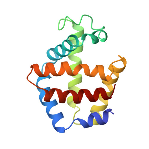
High-resolution crystallographic analysis of a co-operative dimeric hemoglobin.
Royer Jr., W.E.(1994) J Mol Biol 235: 657-681
- PubMed: 8289287
- DOI: https://doi.org/10.1006/jmbi.1994.1019
- Primary Citation of Related Structures:
3SDH, 4SDH - PubMed Abstract:
High-resolution crystal structures of the co-operative dimeric hemoglobin from the blood clam Scapharca inaequivalvis have been determined in the unliganded (deoxy) and carbon monoxide (CO) liganded states. The deoxy structure has been refined at 1.6 A resolution to an R-factor of 0.158 and the CO structure has been refined at 1.4 A resolution to an R-factor of 0.159. These structures reveal details of the structural transitions involved in co-operative ligand binding that involve only a minor rotation of subunits but very striking tertiary changes at the interface. A small number of residues in the F-helix appear to mediate co-operativity in this simple hemoglobin. The oxygen affinity of each subunit appears to be largely dictated by the disposition of phenylalanine 97, whose side-chain packs in the heme pocket in the deoxy state but is extruded towards the interface in the CO-liganded structure. Direct involvement of the ligand-binding heme group is a novel feature of the subunit interface and appears important for intersubunit communication. Ligation alters the conformation of the heme propionate groups along with two interacting residues from the symmetry-related subunit. These two residues, lysine 96 and asparagine 100, link the heme of one subunit with the F-helix of the second subunit in such a way as to influence the ligand affinity of that subunit. The interface is highly hydrated by well-ordered water molecules that are likely to be important in the stabilization of the two structures.
Organizational Affiliation:
Program in Molecular Medicine, University of Massachusetts Medical Center, Worcester 01605.