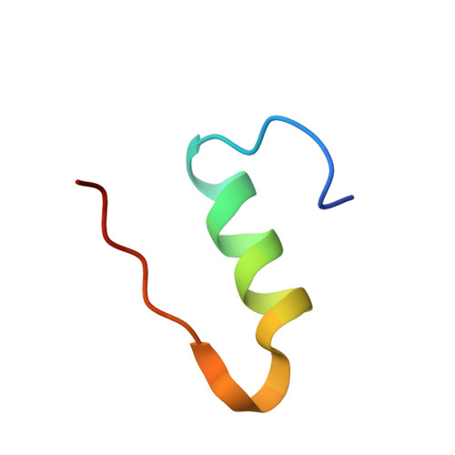
4A7E

X-ray crystal structure of porcine insulin flash-cooled at high pressure


Experimental Data Snapshot

  • Method: X-RAY DIFFRACTION
  • Resolution: 1.86 Å
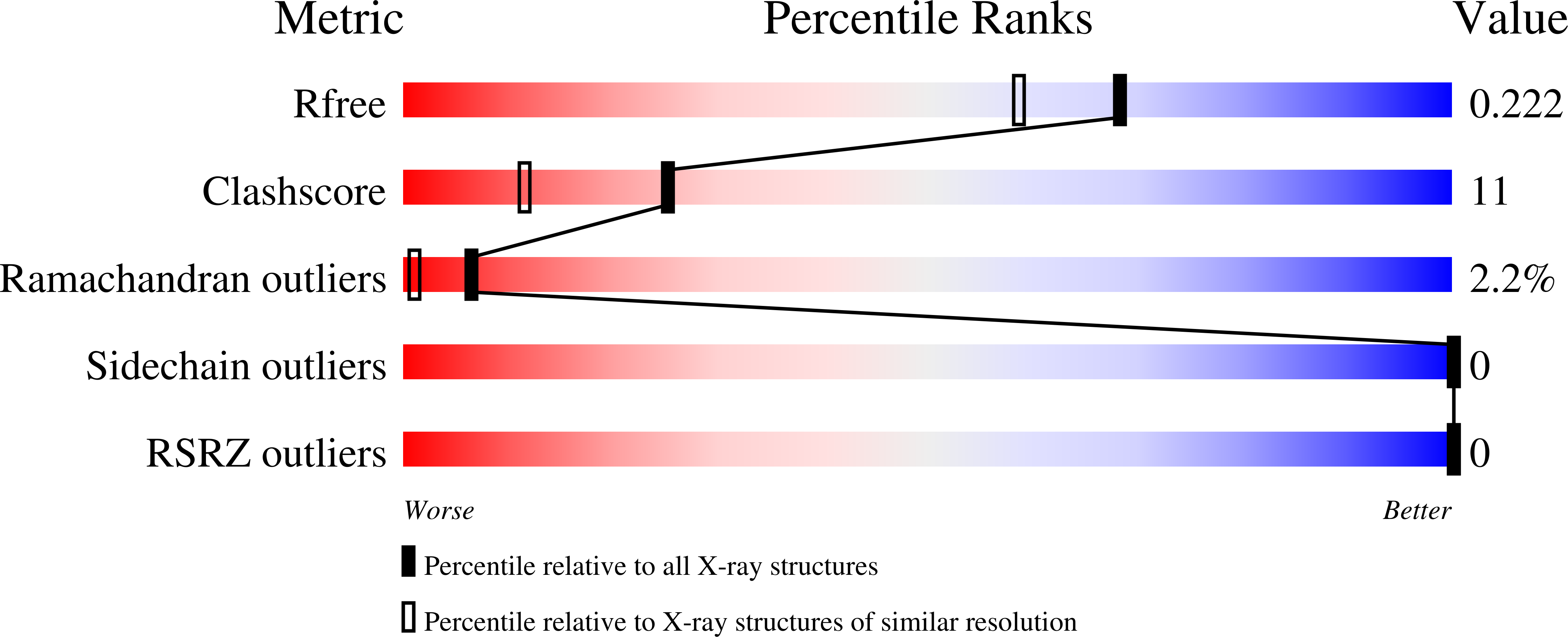
  • R-Value Free: 0.217 
  • R-Value Work: 0.176 
  • R-Value Observed: 0.182 

Starting Model: experimental
View more details

wwPDB Validation   3D Report Full Report


This is version 1.4 of the entry. See complete history


Literature

Fast High-Pressure Freezing of Protein Crystals in Their Mother Liquor

Burkhardt, A.Warmer, M.Pannerselvam, S.Wagner, A.Zouni, A.Reimer, R.Hohenberg, H.Meents, A.

(2012) Acta Crystallogr Sect F Struct Biol Cryst Commun 68: 495

  • DOI: https://doi.org/10.1107/S1744309112009670
  • Primary Citation of Related Structures:  
    4A7D, 4A7E

  • PubMed Abstract: 

    High-pressure freezing (HPF) is a method which allows sample vitrification without cryoprotectants. In the present work, protein crystals were cooled to cryogenic temperatures at a pressure of 210 MPa. In contrast to other HPF methods published to date in the field of cryocrystallography, this protocol involves rapid sample cooling using a standard HPF device. The fast cooling rates allow HPF of protein crystals directly in their mother liquor without the need for cryoprotectants or external reagents. HPF was first attempted with hen egg-white lysozyme and cubic insulin crystals, yielding good to excellent diffraction quality. Non-cryoprotected crystals of the membrane protein photosystem II have been successfully cryocooled for the first time. This indicates that the presented HPF method is well suited to the vitrification of challenging systems with large unit cells and weak crystal contacts.


  • Organizational Affiliation

    Deutsches Elektronen-Synchrotron DESY, Notkestrasse 85, 22607 Hamburg, Germany. anja.burkhardt@desy.de


Macromolecules

Find similar proteins by:  Sequence   |   3D Structure  

Entity ID: 1
MoleculeChains Sequence LengthOrganismDetailsImage
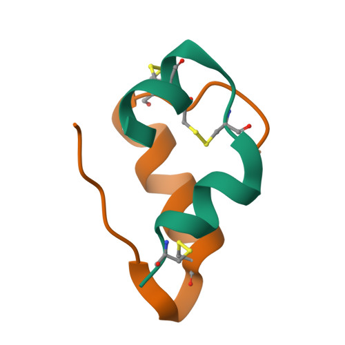
INSULIN A CHAIN21Sus scrofaMutation(s): 0 
UniProt
Find proteins for P01315 (Sus scrofa)
Explore P01315 
Go to UniProtKB:  P01315
Entity Groups  
Sequence Clusters30% Identity50% Identity70% Identity90% Identity95% Identity100% Identity
UniProt GroupP01315
Sequence Annotations
Expand
  • Reference Sequence
Find similar proteins by:  (by identity cutoff)  |  3D Structure
Entity ID: 2
MoleculeChains Sequence LengthOrganismDetailsImage
INSULIN B CHAIN30Sus scrofaMutation(s): 0 
UniProt
Find proteins for P01315 (Sus scrofa)
Explore P01315 
Go to UniProtKB:  P01315
Entity Groups  
Sequence Clusters30% Identity50% Identity70% Identity90% Identity95% Identity100% Identity
UniProt GroupP01315
Sequence Annotations
Expand
  • Reference Sequence
Experimental Data & Validation

Experimental Data

  • Method: X-RAY DIFFRACTION
  • Resolution: 1.86 Å
  • R-Value Free: 0.217 
  • R-Value Work: 0.176 
  • R-Value Observed: 0.182 
  • Space Group: I 21 3
Unit Cell:
Length ( Å )Angle ( ˚ )
a = 77.1α = 90
b = 77.1β = 90
c = 77.1γ = 90
Software Package:
Software NamePurpose
PHENIXrefinement
XDSdata reduction
XSCALEdata scaling
PHENIXphasing

Structure Validation

View Full Validation Report


Entry History 

Deposition Data

Revision History  (Full details and data files)

  • Version 1.0: 2011-11-30
    Type: Initial release
  • Version 1.1: 2012-04-11
    Changes: Other
  • Version 1.2: 2012-05-30
    Changes: Other
  • Version 1.3: 2023-12-20
    Changes: Data collection, Database references, Other, Refinement description
  • Version 1.4: 2024-10-09
    Changes: Structure summary