5OGH

Structure of RNase A at high resolution (1.16 A) in complex with 3'-CMP and sulphate ions


Experimental Data Snapshot

  • Method: X-RAY DIFFRACTION
  • Resolution: 1.16 Å
  • R-Value Free: 0.135 
  • R-Value Work: 0.121 
  • R-Value Observed: 0.121 

wwPDB Validation   3D Report Full Report


Ligand Structure Quality Assessment 


This is version 1.3 of the entry. See complete history


Literature

Characterization of an RNase with two catalytic centers. Human RNase6 catalytic and phosphate-binding site arrangement favors the endonuclease cleavage of polymeric substrates.

Prats-Ejarque, G.Blanco, J.A.Salazar, V.A.Nogues, V.M.Moussaoui, M.Boix, E.

(2019) Biochim Biophys Acta Gen Subj 1863: 105-117

  • DOI: https://doi.org/10.1016/j.bbagen.2018.09.021
  • Primary Citation of Related Structures:  
    5ET4, 5OAB, 5OGH, 6ENP

  • PubMed Abstract: 

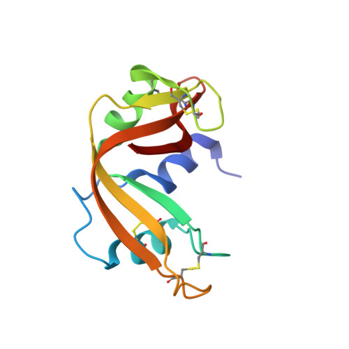
    Human RNase6 is a small cationic antimicrobial protein that belongs to the vertebrate RNaseA superfamily. All members share a common catalytic mechanism, which involves a conserved catalytic triad, constituted by two histidines and a lysine (His15/His122/Lys38 in RNase6 corresponding to His12/His119/Lys41 in RNaseA). Recently, our first crystal structure of human RNase6 identified an additional His pair (His36/His39) and suggested the presence of a secondary active site. In this work we have explored RNase6 and RNaseA subsite architecture by X-ray crystallography, site-directed mutagenesis and kinetic characterization. The analysis of two novel crystal structures of RNase6 in complex with phosphate anions at atomic resolution locates a total of nine binding sites and reveals the contribution of Lys87 to phosphate-binding at the secondary active center. Contribution of the second catalytic triad residues to the enzyme activity is confirmed by mutagenesis. RNase6 catalytic site architecture has been compared with an RNaseA engineered variant where a phosphate-binding subsite is converted into a secondary catalytic center (RNaseA-K7H/R10H). We have identified the residues that participate in RNase6 second catalytic triad (His36/His39/Lys87) and secondary phosphate-binding sites. To note, residues His39 and Lys87 are unique within higher primates. The RNaseA/RNase6 side-by-side comparison correlates the presence of a dual active site in RNase6 with a favored endonuclease-type cleavage pattern. An RNase dual catalytic and extended binding site arrangement facilitates the cleavage of polymeric substrates. This is the first report of the presence of two catalytic centers in a single monomer within the RNaseA superfamily.


  • Organizational Affiliation

    Department of Biochemistry and Molecular Biology, Faculty of Biosciences, Universitat Autònoma de Barcelona, E-08193 Cerdanyola del Vallès, Spain. Electronic address: Guillem.Prats.Ejarque@uab.cat.


Macromolecules
Find similar proteins by:  (by identity cutoff)  |  3D Structure
Entity ID: 1
MoleculeChains Sequence LengthOrganismDetailsImage
Ribonuclease pancreatic124Bos taurusMutation(s): 0 
EC: 3.1.27.5
UniProt
Find proteins for P61823 (Bos taurus)
Explore P61823 
Go to UniProtKB:  P61823
Entity Groups  
Sequence Clusters30% Identity50% Identity70% Identity90% Identity95% Identity100% Identity
UniProt GroupP61823
Sequence Annotations
Expand
  • Reference Sequence
Small Molecules
Ligands 4 Unique
IDChains Name / Formula / InChI Key2D Diagram3D Interactions
C3P (Subject of Investigation/LOI)
Query on C3P

Download Ideal Coordinates CCD File 
B [auth A],
C [auth A]
CYTIDINE-3'-MONOPHOSPHATE
C9 H14 N3 O8 P
UOOOPKANIPLQPU-XVFCMESISA-N
SO4
Query on SO4

Download Ideal Coordinates CCD File 
D [auth A]SULFATE ION
O4 S
QAOWNCQODCNURD-UHFFFAOYSA-L
CL
Query on CL

Download Ideal Coordinates CCD File 
E [auth A],
F [auth A],
G [auth A],
H [auth A],
I [auth A]
CHLORIDE ION
Cl
VEXZGXHMUGYJMC-UHFFFAOYSA-M
NA
Query on NA

Download Ideal Coordinates CCD File 
J [auth A],
K [auth A]
SODIUM ION
Na
FKNQFGJONOIPTF-UHFFFAOYSA-N
Binding Affinity Annotations 
IDSourceBinding Affinity
C3P BindingDB:  5OGH Ki: min: 2.01e+4, max: 1.03e+5 (nM) from 2 assay(s)
Experimental Data & Validation

Experimental Data

  • Method: X-RAY DIFFRACTION
  • Resolution: 1.16 Å
  • R-Value Free: 0.135 
  • R-Value Work: 0.121 
  • R-Value Observed: 0.121 
  • Space Group: P 32 2 1
Unit Cell:
Length ( Å )Angle ( ˚ )
a = 64.06α = 90
b = 64.06β = 90
c = 64.058γ = 120
Software Package:
Software NamePurpose
PHENIXrefinement
XDSdata reduction
XDSdata scaling
PHASERphasing

Structure Validation

View Full Validation Report


Ligand Structure Quality Assessment 


Entry History & Funding Information

Deposition Data


Funding OrganizationLocationGrant Number
MINECOSpainSAF2015-66007-P
Generalitat de CatalunyaSpain2014 SGR 728

Revision History  (Full details and data files)

  • Version 1.0: 2018-08-01
    Type: Initial release
  • Version 1.1: 2018-10-24
    Changes: Data collection, Database references
  • Version 1.2: 2019-09-11
    Changes: Data collection, Database references
  • Version 1.3: 2024-01-17
    Changes: Advisory, Data collection, Database references, Derived calculations, Refinement description