6IV4

Crystal structure of a bacterial Bestrophin homolog from Klebsiella pneumoniae with a mutation W252F


Experimental Data Snapshot

  • Method: X-RAY DIFFRACTION
  • Resolution: 3.14 Å
  • R-Value Free: 0.295 
  • R-Value Work: 0.237 
  • R-Value Observed: 0.243 

wwPDB Validation   3D Report Full Report


This is version 1.1 of the entry. See complete history


Literature

Dual Ca2+-dependent gates in human Bestrophin1 underlie disease-causing mechanisms of gain-of-function mutations.

Ji, C.Kittredge, A.Hopiavuori, A.Ward, N.Chen, S.Fukuda, Y.Zhang, Y.Yang, T.

(2019) Commun Biol 2: 240-240

  • DOI: https://doi.org/10.1038/s42003-019-0433-3
  • Primary Citation of Related Structures:  
    6IV0, 6IV1, 6IV2, 6IV3, 6IV4, 6IVJ, 6IVK, 6IVL, 6IVM, 6IVN, 6IVO, 6IVP, 6IVQ, 6IVR, 6IVW, 6JLF

  • PubMed Abstract: 

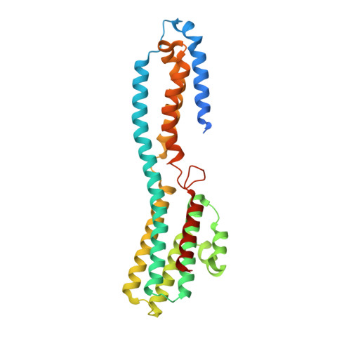
    Mutations of human BEST1 , encoding a Ca 2+ -activated Cl - channel (hBest1), cause macular degenerative disorders. Best1 homolog structures reveal an evolutionarily conserved channel architecture highlighted by two landmark restrictions (named the "neck" and "aperture", respectively) in the ion conducting pathway, suggesting a unique dual-switch gating mechanism, which, however, has not been characterized well. Using patch clamp and crystallography, we demonstrate that both the neck and aperture in hBest1 are Ca 2+ -dependent gates essential for preventing channel leakage resulting from Ca 2+ -independent, spontaneous gate opening. Importantly, three patient-derived mutations (D203A, I205T and Y236C) lead to Ca 2+ -independent leakage and elevated Ca 2+ -dependent anion currents due to enhanced opening of the gates. Moreover, we identify a network of residues critically involved in gate operation. Together, our results suggest an indispensable role of the neck and aperture of hBest1 for channel gating, and uncover disease-causing mechanisms of hBest1 gain-of-function mutations.


  • Organizational Affiliation

    Department of Pharmacology and Physiology, University of Rochester, School of Medicine and Dentistry, Rochester, NY 14642 USA.


Macromolecules
Find similar proteins by:  (by identity cutoff)  |  3D Structure
Entity ID: 1
MoleculeChains Sequence LengthOrganismDetailsImage
Bestrophin homolog
A, B, C, D, E
296Klebsiella pneumoniae IS53Mutation(s): 1 
Entity Groups  
Sequence Clusters30% Identity50% Identity70% Identity90% Identity95% Identity100% Identity
Sequence Annotations
Expand
  • Reference Sequence
Small Molecules
Ligands 1 Unique
IDChains Name / Formula / InChI Key2D Diagram3D Interactions
ZN
Query on ZN

Download Ideal Coordinates CCD File 
F [auth A]
G [auth A]
H [auth A]
I [auth B]
J [auth B]
F [auth A],
G [auth A],
H [auth A],
I [auth B],
J [auth B],
K [auth C],
L [auth C],
M [auth C],
N [auth D],
O [auth D],
P [auth D],
Q [auth E],
R [auth E]
ZINC ION
Zn
PTFCDOFLOPIGGS-UHFFFAOYSA-N
Experimental Data & Validation

Experimental Data

  • Method: X-RAY DIFFRACTION
  • Resolution: 3.14 Å
  • R-Value Free: 0.295 
  • R-Value Work: 0.237 
  • R-Value Observed: 0.243 
  • Space Group: P 21 21 21
Unit Cell:
Length ( Å )Angle ( ˚ )
a = 104.009α = 90
b = 154.186β = 90
c = 161.736γ = 90
Software Package:
Software NamePurpose
PHENIXrefinement
XDSdata reduction
Aimlessdata scaling
PHASERphasing

Structure Validation

View Full Validation Report


Entry History & Funding Information

Deposition Data


Funding OrganizationLocationGrant Number
National Natural Science Foundation of ChinaChina31770801

Revision History  (Full details and data files)

  • Version 1.0: 2019-11-13
    Type: Initial release
  • Version 1.1: 2022-06-08
    Changes: Database references, Derived calculations, Structure summary