


Expression, crystallization, and preliminary X-ray crystallographic analysis of peptide deformylase from Acinetobacter baumanii
Kang, L.W.(2017) Korean Soc Struct Biology 5: 49-52
Experimental Data Snapshot
wwPDB Validation 3D Report Full Report

(2017) Korean Soc Struct Biology 5: 49-52
Entity ID: 1 | |||||
|---|---|---|---|---|---|
| Molecule | Chains | Sequence Length | Organism | Details | Image |
| Peptide deformylase | 170 | Acinetobacter baumannii MRSN 3527 | Mutation(s): 0 Gene Names: def_1, def, T630_0214 EC: 3.5.1.88 | 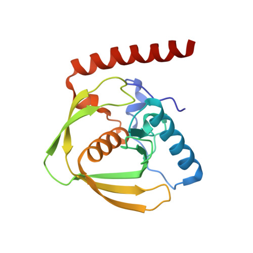 | |
UniProt | |||||
Find proteins for B0VNL8 (Acinetobacter baumannii (strain SDF)) Explore B0VNL8 Go to UniProtKB: B0VNL8 | |||||
Entity Groups | |||||
| Sequence Clusters | 30% Identity50% Identity70% Identity90% Identity95% Identity100% Identity | ||||
| UniProt Group | B0VNL8 | ||||
Sequence AnnotationsExpand | |||||
| |||||
| Ligands 1 Unique | |||||
|---|---|---|---|---|---|
| ID | Chains | Name / Formula / InChI Key | 2D Diagram | 3D Interactions | |
| ZN Query on ZN | C [auth A], D [auth B] | ZINC ION Zn PTFCDOFLOPIGGS-UHFFFAOYSA-N |  | ||
| Length ( Å ) | Angle ( ˚ ) |
|---|---|
| a = 39.425 | α = 90 |
| b = 39.425 | β = 90 |
| c = 187.888 | γ = 120 |
| Software Name | Purpose |
|---|---|
| REFMAC | refinement |
| HKL-2000 | data scaling |
| HKL-2000 | data collection |
| HKL-2000 | data reduction |
| MOLREP | phasing |
RCSB PDB (citation) is hosted by
RCSB PDB is a member of the

