6OAH

Crystal structure of human FPPS in complex with an allosteric inhibitor YF-02-78


Experimental Data Snapshot

  • Method: X-RAY DIFFRACTION
  • Resolution: 2.20 Å
  • R-Value Free: 0.260 
  • R-Value Work: 0.208 
  • R-Value Observed: 0.210 

wwPDB Validation   3D Report Full Report


Ligand Structure Quality Assessment 


This is version 1.3 of the entry. See complete history


Literature

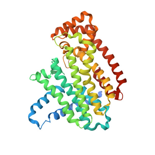
Chirality-Driven Mode of Binding of alpha-Aminophosphonic Acid-Based Allosteric Inhibitors of the Human Farnesyl Pyrophosphate Synthase (hFPPS).

Feng, Y.Park, J.Li, S.G.Boutin, R.Viereck, P.Schilling, M.A.Berghuis, A.M.Tsantrizos, Y.S.

(2019) J Med Chem 62: 9691-9702

  • DOI: https://doi.org/10.1021/acs.jmedchem.9b01104
  • Primary Citation of Related Structures:  
    6N7Y, 6N7Z, 6N82, 6N83, 6OAG, 6OAH

  • PubMed Abstract: 

    Thienopyrimidine-based allosteric inhibitors of the human farnesyl pyrophosphate synthase (hFPPS), characterized by a chiral α-aminophosphonic acid moiety, were synthesized as enantiomerically enriched pairs, and their binding mode was investigated by X-ray crystallography. A general consensus in the binding orientation of all ( R )- and ( S )-enantiomers was revealed. This finding is a prerequisite for establishing a reliable structure-activity relationship (SAR) model.


  • Organizational Affiliation

    Department of Chemistry , McGill University , 801 Sherbrooke Street West , Montreal , Quebec H3A 0B8 , Canada.


Macromolecules
Find similar proteins by:  (by identity cutoff)  |  3D Structure
Entity ID: 1
MoleculeChains Sequence LengthOrganismDetailsImage
Farnesyl pyrophosphate synthaseA [auth F]375Homo sapiensMutation(s): 0 
Gene Names: FDPSFPSKIAA1293
EC: 2.5.1.10 (PDB Primary Data), 2.5.1.1 (PDB Primary Data)
UniProt & NIH Common Fund Data Resources
Find proteins for P14324 (Homo sapiens)
Explore P14324 
Go to UniProtKB:  P14324
PHAROS:  P14324
GTEx:  ENSG00000160752 
Entity Groups  
Sequence Clusters30% Identity50% Identity70% Identity90% Identity95% Identity100% Identity
UniProt GroupP14324
Sequence Annotations
Expand
  • Reference Sequence
Small Molecules
Binding Affinity Annotations 
IDSourceBinding Affinity
M2V Binding MOAD:  6OAH IC50: 1500 (nM) from 1 assay(s)
Experimental Data & Validation

Experimental Data

  • Method: X-RAY DIFFRACTION
  • Resolution: 2.20 Å
  • R-Value Free: 0.260 
  • R-Value Work: 0.208 
  • R-Value Observed: 0.210 
  • Space Group: P 41 21 2
Unit Cell:
Length ( Å )Angle ( ˚ )
a = 111.33α = 90
b = 111.33β = 90
c = 70γ = 90
Software Package:
Software NamePurpose
REFMACrefinement
XDSdata reduction
XSCALEdata scaling
REFMACphasing

Structure Validation

View Full Validation Report


Ligand Structure Quality Assessment 


Entry History & Funding Information

Deposition Data


Funding OrganizationLocationGrant Number
Canadian Institutes of Health Research (CIHR)Canada--

Revision History  (Full details and data files)

  • Version 1.0: 2019-11-06
    Type: Initial release
  • Version 1.1: 2019-11-27
    Changes: Database references
  • Version 1.2: 2020-01-08
    Changes: Author supporting evidence
  • Version 1.3: 2023-10-11
    Changes: Data collection, Database references, Refinement description