6TF1

Crystal structure of the ADP-binding domain of the NAD+ riboswitch with Adenosine diphosphate (ADP)


Experimental Data Snapshot

  • Method: X-RAY DIFFRACTION
  • Resolution: 2.40 Å
  • R-Value Free: 0.245 
  • R-Value Work: 0.216 
  • R-Value Observed: 0.217 

wwPDB Validation   3D Report Full Report


Ligand Structure Quality Assessment 


This is version 1.1 of the entry. See complete history


Literature

Structure and ligand binding of the ADP-binding domain of the NAD+ riboswitch.

Huang, L.Wang, J.Lilley, D.M.J.

(2020) RNA 26: 878-887

  • DOI: https://doi.org/10.1261/rna.074898.120
  • Primary Citation of Related Structures:  
    6TB7, 6TF0, 6TF1, 6TF2, 6TF3, 6TFE, 6TFF, 6TFG, 6TFH

  • PubMed Abstract: 

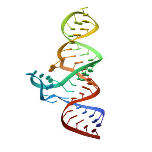
    The nad A motif is the first known NAD + -dependent riboswitch, comprising two similar tandem bulged stem-loop structures. We have determined the structure of the 5' domain 1 of the riboswitch. It has three coaxial helical segments, separated by an ACANCCCC bulge and by an internal loop, with a tertiary contact between them that includes two C:G base pairs. We have determined the structure with a number of ligands related to NADH, but in each case only the ADP moiety is observed. The adenosine adopts an anti conformation, forms multiple hydrogen bonds across the width of the sugar edge of the penultimate C:G base pair of the helix preceding the bulge, and the observed contacts have been confirmed by mutagenesis and calorimetry. Two divalent metal ions play a key structural role at the narrow neck of the bulge. One makes direct bonding contacts to the diphosphate moiety, locking it into position. Thus the nucleobase, ribose, and phosphate groups of the ADP moiety are all specifically recognized by the RNA. The NAD + riboswitch is modular. Domain 1 is an ADP binding domain that may be ancient and could potentially be used in combination with other ligand binding motifs such as CoA.


  • Organizational Affiliation

    Cancer Research UK Nucleic Acid Structure Research Group, MSI/WTB Complex, The University of Dundee, Dundee DD1 5EH, United Kingdom.


Macromolecules
Find similar nucleic acids by:  (by identity cutoff)  |  3D Structure
Entity ID: 1
MoleculeChains LengthOrganismImage
Chains: A52Candidatus Koribacter versatilis Ellin345
Sequence Annotations
Expand
  • Reference Sequence
Small Molecules
Ligands 3 Unique
IDChains Name / Formula / InChI Key2D Diagram3D Interactions
ADP (Subject of Investigation/LOI)
Query on ADP

Download Ideal Coordinates CCD File 
J [auth A]ADENOSINE-5'-DIPHOSPHATE
C10 H15 N5 O10 P2
XTWYTFMLZFPYCI-KQYNXXCUSA-N
MG
Query on MG

Download Ideal Coordinates CCD File 
B [auth A]
C [auth A]
D [auth A]
E [auth A]
F [auth A]
B [auth A],
C [auth A],
D [auth A],
E [auth A],
F [auth A],
G [auth A],
H [auth A]
MAGNESIUM ION
Mg
JLVVSXFLKOJNIY-UHFFFAOYSA-N
NA
Query on NA

Download Ideal Coordinates CCD File 
I [auth A]SODIUM ION
Na
FKNQFGJONOIPTF-UHFFFAOYSA-N
Experimental Data & Validation

Experimental Data

  • Method: X-RAY DIFFRACTION
  • Resolution: 2.40 Å
  • R-Value Free: 0.245 
  • R-Value Work: 0.216 
  • R-Value Observed: 0.217 
  • Space Group: I 2 2 2
Unit Cell:
Length ( Å )Angle ( ˚ )
a = 57.585α = 90
b = 59.014β = 90
c = 190.587γ = 90
Software Package:
Software NamePurpose
PHENIXrefinement
PHENIXrefinement
xia2data reduction
xia2data scaling
PHASERphasing

Structure Validation

View Full Validation Report


Ligand Structure Quality Assessment 


Entry History & Funding Information

Deposition Data


Funding OrganizationLocationGrant Number
Cancer Research UKUnited KingdomA18604

Revision History  (Full details and data files)

  • Version 1.0: 2020-09-23
    Type: Initial release
  • Version 1.1: 2024-01-24
    Changes: Data collection, Database references, Refinement description