


6U7H
Cryo-EM structure of the HCoV-229E spike glycoprotein
- PDB DOI: https://doi.org/10.2210/pdb6U7H/pdb
- EM Map EMD-20668: EMDB EMDataResource
- Classification: VIRAL PROTEIN
- Organism(s): Human coronavirus 229E
- Expression System: Homo sapiens
- Mutation(s): No
- Deposited: 2019-09-02 Released: 2019-11-13
- Funding Organization(s): Canadian Institutes of Health Research (CIHR)
Experimental Data Snapshot
- Method: ELECTRON MICROSCOPY
- Resolution: 3.10 Å
- Aggregation State: PARTICLE
- Reconstruction Method: SINGLE PARTICLE
wwPDB Validation 3D Report Full Report

This is version 2.0 of the entry. See complete history.

Macromolecules
Find similar proteins by:
(by identity cutoff) | 3D Structure
Entity ID: 1 | |||||
|---|---|---|---|---|---|
| Molecule | Chains | Sequence Length | Organism | Details | Image |
| spike glycoprotein | 1,159 | Human coronavirus 229E | Mutation(s): 0 | 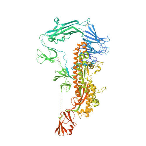 | |
UniProt | |||||
Find proteins for P15423 (Human coronavirus 229E) Explore P15423 Go to UniProtKB: P15423 | |||||
Entity Groups | |||||
| Sequence Clusters | 30% Identity50% Identity70% Identity90% Identity95% Identity100% Identity | ||||
| UniProt Group | P15423 | ||||
Sequence AnnotationsExpand | |||||
| |||||
Oligosaccharides
Entity ID: 2 | |||||
|---|---|---|---|---|---|
| Molecule | Chains | Length | 2D Diagram | Glycosylation | 3D Interactions |
| alpha-D-mannopyranose-(1-2)-alpha-D-mannopyranose-(1-3)-[alpha-D-mannopyranose-(1-3)-alpha-D-mannopyranose-(1-6)]beta-D-mannopyranose-(1-4)-2-acetamido-2-deoxy-beta-D-glucopyranose-(1-4)-2-acetamido-2-deoxy-beta-D-glucopyranose | D, I, N | 7 |  | N-Glycosylation | |
Glycosylation Resources | |||||
GlyTouCan: G07617FP GlyCosmos: G07617FP GlyGen: G07617FP | |||||


Small Molecules
| Ligands 1 Unique | |||||
|---|---|---|---|---|---|
| ID | Chains | Name / Formula / InChI Key | 2D Diagram | 3D Interactions | |
| NAG Query on NAG | AA [auth A] BA [auth A] CA [auth A] DA [auth B] EA [auth B] | 2-acetamido-2-deoxy-beta-D-glucopyranose C8 H15 N O6 OVRNDRQMDRJTHS-FMDGEEDCSA-N |  | ||
Experimental Data & Validation
Experimental Data
- Method: ELECTRON MICROSCOPY
- Resolution: 3.10 Å
- Aggregation State: PARTICLE
- Reconstruction Method: SINGLE PARTICLE
| Task | Software Package | Version |
|---|---|---|
| RECONSTRUCTION | cryoSPARC | 2.90 |
| MODEL REFINEMENT | PHENIX | 1.16RC1-3535 |
Entry History & Funding Information
Deposition Data
- Released Date: 2019-11-13 Deposition Author(s): Li, Z., Benlekbir, S., Rubinstein, J.L., Rini, J.M.
| Funding Organization | Location | Grant Number |
|---|---|---|
| Canadian Institutes of Health Research (CIHR) | Canada | -- |
Revision History (Full details and data files)
- Version 1.0: 2019-11-13
Type: Initial release - Version 1.1: 2020-01-15
Changes: Author supporting evidence - Version 2.0: 2020-07-29
Type: Remediation
Reason: Carbohydrate remediation
Changes: Atomic model, Data collection, Derived calculations, Structure summary
