6WZ5

Bridging of double-strand DNA break activates PARP2/HPF1 to modify chromatin


Experimental Data Snapshot

  • Method: ELECTRON MICROSCOPY
  • Resolution: 2.20 Å
  • Aggregation State: PARTICLE 
  • Reconstruction Method: SINGLE PARTICLE 

wwPDB Validation   3D Report Full Report


This is version 1.3 of the entry. See complete history


Literature

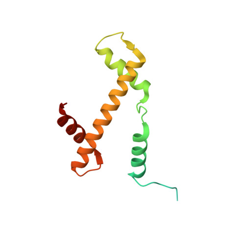
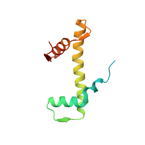
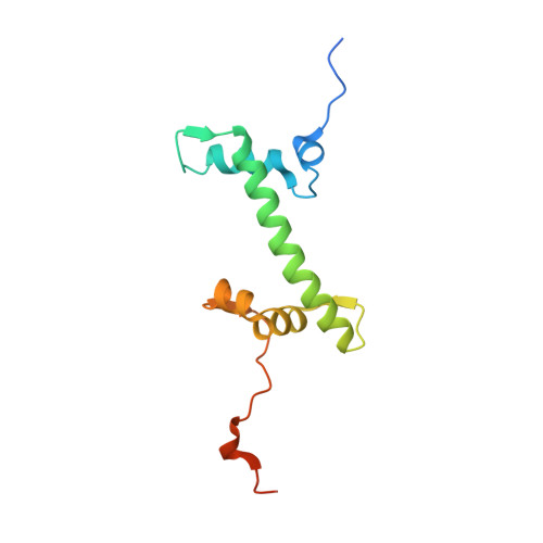
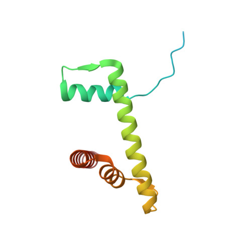
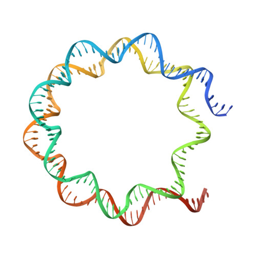
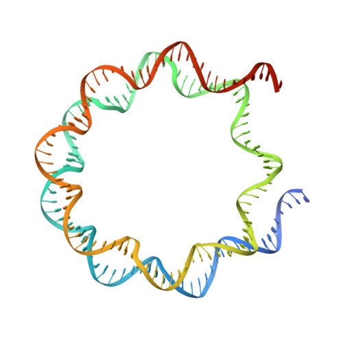
Bridging of DNA breaks activates PARP2-HPF1 to modify chromatin.

Bilokapic, S.Suskiewicz, M.J.Ahel, I.Halic, M.

(2020) Nature 585: 609-613

  • DOI: https://doi.org/10.1038/s41586-020-2725-7
  • Primary Citation of Related Structures:  
    6WZ5, 6WZ9, 6X0L, 6X0M, 6X0N

  • PubMed Abstract: 

    Breaks in DNA strands recruit the protein PARP1 and its paralogue PARP2 to modify histones and other substrates through the addition of mono- and poly(ADP-ribose) (PAR) 1-5 . In the DNA damage responses, this post-translational modification occurs predominantly on serine residues 6-8 and requires HPF1, an accessory factor that switches the amino acid specificity of PARP1 and PARP2 from aspartate or glutamate to serine 9,10 . Poly(ADP) ribosylation (PARylation) is important for subsequent chromatin decompaction and provides an anchor for the recruitment of downstream signalling and repair factors to the sites of DNA breaks 2,11 . Here, to understand the molecular mechanism by which PARP enzymes recognize DNA breaks within chromatin, we determined the cryo-electron-microscopic structure of human PARP2-HPF1 bound to a nucleosome. This showed that PARP2-HPF1 bridges two nucleosomes, with the broken DNA aligned in a position suitable for ligation, revealing the initial step in the repair of double-strand DNA breaks. The bridging induces structural changes in PARP2 that signal the recognition of a DNA break to the catalytic domain, which licenses HPF1 binding and PARP2 activation. Our data suggest that active PARP2 cycles through different conformational states to exchange NAD + and substrate, which may enable PARP enzymes to act processively while bound to chromatin. The processes of PARP activation and the PARP catalytic cycle we describe can explain mechanisms of resistance to PARP inhibitors and will aid the development of better inhibitors as cancer treatments 12-16 .


  • Organizational Affiliation

    Department of Structural Biology, St Jude Children's Research Hospital, Memphis, TN, USA.


Macromolecules

Find similar proteins by:  (by identity cutoff)  |  3D Structure
Entity ID: 1
MoleculeChains Sequence LengthOrganismDetailsImage
Histone H3.2
A, E
135Xenopus laevisMutation(s): 1 
UniProt
Find proteins for P84233 (Xenopus laevis)
Explore P84233 
Go to UniProtKB:  P84233
Entity Groups  
Sequence Clusters30% Identity50% Identity70% Identity90% Identity95% Identity100% Identity
UniProt GroupP84233
Sequence Annotations
Expand
  • Reference Sequence
Find similar proteins by:  (by identity cutoff)  |  3D Structure
Entity ID: 2
MoleculeChains Sequence LengthOrganismDetailsImage
Histone H4
B, F
102Xenopus laevisMutation(s): 0 
UniProt
Find proteins for P62799 (Xenopus laevis)
Explore P62799 
Go to UniProtKB:  P62799
Entity Groups  
Sequence Clusters30% Identity50% Identity70% Identity90% Identity95% Identity100% Identity
UniProt GroupP62799
Sequence Annotations
Expand
  • Reference Sequence
Find similar proteins by:  (by identity cutoff)  |  3D Structure
Entity ID: 3
MoleculeChains Sequence LengthOrganismDetailsImage
Histone H2A
C, G
129Xenopus laevisMutation(s): 0 
Gene Names: hist1h2ajh2ac14LOC494591XELAEV_18003602mg
UniProt
Find proteins for P06897 (Xenopus laevis)
Explore P06897 
Go to UniProtKB:  P06897
Entity Groups  
Sequence Clusters30% Identity50% Identity70% Identity90% Identity95% Identity100% Identity
UniProt GroupP06897
Sequence Annotations
Expand
  • Reference Sequence
Find similar proteins by:  (by identity cutoff)  |  3D Structure
Entity ID: 4
MoleculeChains Sequence LengthOrganismDetailsImage
Histone H2B 1.1
D, H
122Xenopus laevisMutation(s): 1 
UniProt
Find proteins for P02281 (Xenopus laevis)
Explore P02281 
Go to UniProtKB:  P02281
Entity Groups  
Sequence Clusters30% Identity50% Identity70% Identity90% Identity95% Identity100% Identity
UniProt GroupP02281
Sequence Annotations
Expand
  • Reference Sequence
Find similar nucleic acids by:  (by identity cutoff)  |  3D Structure
Entity ID: 5
MoleculeChains LengthOrganismImage
DNA (153-MER)167synthetic construct
Sequence Annotations
Expand
  • Reference Sequence
Find similar nucleic acids by:  (by identity cutoff)  |  3D Structure
Entity ID: 6
MoleculeChains LengthOrganismImage
DNA (153-MER)167synthetic construct
Sequence Annotations
Expand
  • Reference Sequence
Experimental Data & Validation

Experimental Data

  • Method: ELECTRON MICROSCOPY
  • Resolution: 2.20 Å
  • Aggregation State: PARTICLE 
  • Reconstruction Method: SINGLE PARTICLE 

Structure Validation

View Full Validation Report


Entry History & Funding Information

Deposition Data


Funding OrganizationLocationGrant Number
National Institutes of Health/National Institute of General Medical Sciences (NIH/NIGMS)United States1R01GM135599-01

Revision History  (Full details and data files)

  • Version 1.0: 2020-09-16
    Type: Initial release
  • Version 1.1: 2020-09-30
    Changes: Database references
  • Version 1.2: 2020-10-07
    Changes: Database references
  • Version 1.3: 2024-03-06
    Changes: Data collection, Database references