


6YDE
X-ray structure of LPMO
- PDB DOI: https://doi.org/10.2210/pdb6YDE/pdb
- Classification: METAL BINDING PROTEIN
- Organism(s): Achaetomiella virescens
- Expression System: Aspergillus oryzae
- Mutation(s): No
- Deposited: 2020-03-20 Released: 2020-09-16
- Funding Organization(s): Novo Nordisk Foundation
Experimental Data Snapshot
- Method: X-RAY DIFFRACTION
- Resolution: 2.20 Å
- R-Value Free: 0.264
- R-Value Work: 0.194
- R-Value Observed: 0.198
wwPDB Validation 3D Report Full Report

This is version 1.2 of the entry. See complete history.

Macromolecules
Find similar proteins by:
(by identity cutoff) | 3D Structure
Entity ID: 1 | |||||
|---|---|---|---|---|---|
| Molecule | Chains | Sequence Length | Organism | Details | Image |
| LPMO lytic polysaccharide monooxygenase | 252 | Achaetomiella virescens | Mutation(s): 0 Gene Names: aa9 | 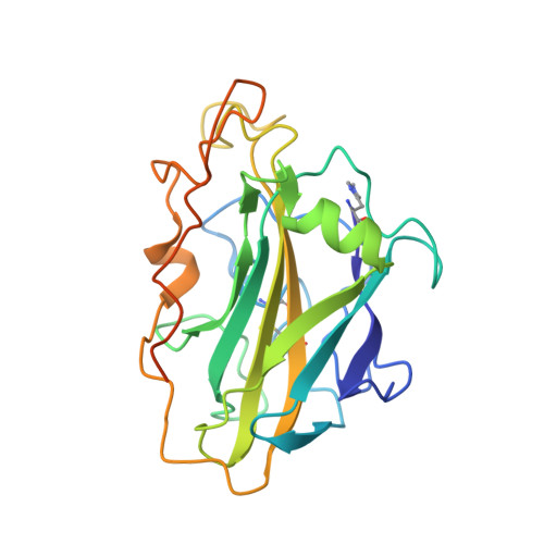 | |
UniProt | |||||
Find proteins for A0A223GEC9 (Collariella virescens) Explore A0A223GEC9 Go to UniProtKB: A0A223GEC9 | |||||
Entity Groups | |||||
| Sequence Clusters | 30% Identity50% Identity70% Identity90% Identity95% Identity100% Identity | ||||
| UniProt Group | A0A223GEC9 | ||||
Sequence AnnotationsExpand | |||||
| |||||
Oligosaccharides
Entity ID: 2 | |||||
|---|---|---|---|---|---|
| Molecule | Chains | Length | 2D Diagram | Glycosylation | 3D Interactions |
| beta-D-glucopyranose-(1-4)-beta-D-glucopyranose-(1-4)-beta-D-glucopyranose-(1-4)-beta-D-glucopyranose-(1-4)-beta-D-glucopyranose-(1-4)-beta-D-glucopyranose | B | 6 |  | N/A | |
Glycosylation Resources | |||||
GlyTouCan: G09454VW GlyCosmos: G09454VW GlyGen: G09454VW | |||||
Small Molecules
| Ligands 2 Unique | |||||
|---|---|---|---|---|---|
| ID | Chains | Name / Formula / InChI Key | 2D Diagram | 3D Interactions | |
| SO4 Query on SO4 | C [auth A] | SULFATE ION O4 S QAOWNCQODCNURD-UHFFFAOYSA-L |  | ||
| CU (Subject of Investigation/LOI) Query on CU | D [auth A] | COPPER (II) ION Cu JPVYNHNXODAKFH-UHFFFAOYSA-N |  | ||
| Modified Residues 1 Unique | |||||
|---|---|---|---|---|---|
| ID | Chains | Type | Formula | 2D Diagram | Parent |
| HIC Query on HIC | A | L-PEPTIDE LINKING | C7 H11 N3 O2 |  | HIS |
Biologically Interesting Molecules (External Reference) 1 Unique
Entity ID: 2 | |||||
|---|---|---|---|---|---|
| ID | Chains | Name | Type/Class | 2D Diagram | 3D Interactions |
| PRD_900020 Query on PRD_900020 | B | beta-cellohexaose | Oligosaccharide / Metabolism |  | |
Experimental Data & Validation
Experimental Data
- Method: X-RAY DIFFRACTION
- Resolution: 2.20 Å
- R-Value Free: 0.264
- R-Value Work: 0.194
- R-Value Observed: 0.198
- Space Group: P 21 21 2
Unit Cell:
| Length ( Å ) | Angle ( ˚ ) |
|---|---|
| a = 39.55 | α = 90 |
| b = 124.37 | β = 90 |
| c = 51.67 | γ = 90 |
| Software Name | Purpose |
|---|---|
| REFMAC | refinement |
| PDB_EXTRACT | data extraction |
| XDS | data reduction |
| XSCALE | data scaling |
| MOLREP | phasing |
Entry History & Funding Information
Deposition Data
- Released Date: 2020-09-16 Deposition Author(s): Tandrup, T., Tryfona, T., Frandsen, K.E.H., Johansen, K.S., Dupree, P., Lo Leggio, L.
| Funding Organization | Location | Grant Number |
|---|---|---|
| Novo Nordisk Foundation | Denmark | NNF17SA0027704 |
Revision History (Full details and data files)
- Version 1.0: 2020-09-16
Type: Initial release - Version 1.1: 2020-09-23
Changes: Database references - Version 1.2: 2024-01-24
Changes: Data collection, Database references, Refinement description
