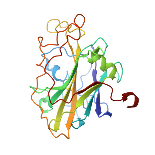
Oligosaccharide Binding and Thermostability of Two Related AA9 Lytic Polysaccharide Monooxygenases.
Tandrup, T., Tryfona, T., Frandsen, K.E.H., Johansen, K.S., Dupree, P., Lo Leggio, L.(2020) Biochemistry 59: 3347-3358
- PubMed: 32818374
- DOI: https://doi.org/10.1021/acs.biochem.0c00312
- Primary Citation of Related Structures:
6YDC, 6YDD, 6YDE, 6YDF, 6YDG - PubMed Abstract:
Lytic polysaccharide monooxygenases (LPMOs) are copper-dependent enzymes that cleave polysaccharide substrates oxidatively. First discovered because of their action on recalcitrant crystalline substrates (chitin and cellulose), a number of LPMOs are now reported to act on soluble substrates, including oligosaccharides. However, crystallographic complexes with oligosaccharides have been reported for only a single LPMO so far, an enzyme from the basidiomycete fungus Lentinus similis ( Ls AA9_A). Here we present a more detailed comparative study of Ls AA9_A and an LPMO from the ascomycete fungus Collariella virescens ( Cv AA9_A) with which it shares 41.5% sequence identity. Ls AA9_A is considerably more thermostable than Cv AA9_A, and the structural basis for the difference has been investigated. We have compared the patterns of oligosaccharide cleavage and the patterns of binding in several new crystal structures explaining the basis for the product preferences of the two enzymes. Obtaining structural information about complexes of LPMOs with carbohydrates has proven to be very difficult in general judging from the structures reported in the literature thus far, and this can be attributed only partly to the low affinity for small substrates. We have thus evaluated the use of differential scanning fluorimetry as a guide to obtaining complex structures. Furthermore, an analysis of crystal packing of LPMOs and glycoside hydrolases corroborates the hypothesis that active site occlusion is a very significant problem for LPMO-substrate interaction analysis by crystallography, due to their relatively flat and extended substrate binding sites.
Organizational Affiliation:
Department of Chemistry, University of Copenhagen, Universitetsparken 5, 2100-DK Copenhagen, Denmark.