6PZW

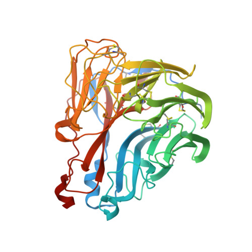
CryoEM derived model of NA-22 Fab in complex with N9 Shanghai2


Experimental Data Snapshot

  • Method: ELECTRON MICROSCOPY
  • Resolution: 3.00 Å
  • Aggregation State: PARTICLE 
  • Reconstruction Method: SINGLE PARTICLE 

Starting Model: experimental
View more details

wwPDB Validation   3D Report Full Report


This is version 2.2 of the entry. See complete history


Literature

Structural Basis of Protection against H7N9 Influenza Virus by Human Anti-N9 Neuraminidase Antibodies.

Zhu, X.Turner, H.L.Lang, S.McBride, R.Bangaru, S.Gilchuk, I.M.Yu, W.Paulson, J.C.Crowe Jr., J.E.Ward, A.B.Wilson, I.A.

(2019) Cell Host Microbe 26: 729

  • DOI: https://doi.org/10.1016/j.chom.2019.10.002
  • Primary Citation of Related Structures:  
    6PZD, 6PZE, 6PZF, 6PZG, 6PZH, 6PZW, 6PZY, 6PZZ, 6U02

  • PubMed Abstract: 

    Influenza virus neuraminidase (NA) is a major target for small-molecule antiviral drugs. Antibodies targeting the NA surface antigen could also inhibit virus entry and egress to provide host protection. However, our understanding of the nature and range of target epitopes is limited because of a lack of human antibody structures with influenza neuraminidase. Here, we describe crystal and cryogenic electron microscopy (cryo-EM) structures of NAs from human-infecting avian H7N9 viruses in complex with five human anti-N9 antibodies, systematically defining several antigenic sites and antibody epitope footprints. These antibodies either fully or partially block the NA active site or bind to epitopes distant from the active site while still showing neuraminidase inhibition. The inhibition of antibodies to NAs was further analyzed by glycan array and solution-based NA activity assays. Together, these structural studies provide insights into protection by anti-NA antibodies and templates for the development of NA-based influenza virus vaccines and therapeutics.


  • Organizational Affiliation

    Department of Integrative Structural and Computational Biology, The Scripps Research Institute, La Jolla, CA 92037, USA.


Macromolecules
Find similar proteins by:  (by identity cutoff)  |  3D Structure
Entity ID: 1
MoleculeChains Sequence LengthOrganismDetailsImage
NeuraminidaseA,
D,
I [auth C],
J [auth B]
429Influenza A virus (A/environment/Shanghai/S1439/2013(H7N9))Mutation(s): 0 
Gene Names: NA
EC: 3.2.1.18
UniProt
Find proteins for S5MF06 (Influenza A virus)
Explore S5MF06 
Go to UniProtKB:  S5MF06
Entity Groups  
Sequence Clusters30% Identity50% Identity70% Identity90% Identity95% Identity100% Identity
UniProt GroupS5MF06
Glycosylation
Glycosylation Sites: 2
Sequence Annotations
Expand
  • Reference Sequence
Find similar proteins by:  (by identity cutoff)  |  3D Structure
Entity ID: 2
MoleculeChains Sequence LengthOrganismDetailsImage
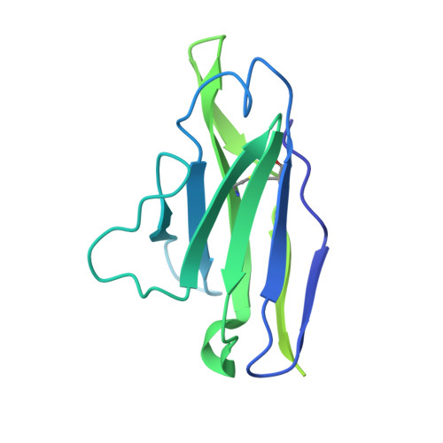
NA-22 fragment antigen binding light chainB [auth I],
E,
G,
K [auth L]
216Homo sapiensMutation(s): 0 
Entity Groups  
Sequence Clusters30% Identity50% Identity70% Identity90% Identity95% Identity100% Identity
Sequence Annotations
Expand
  • Reference Sequence
Find similar proteins by:  (by identity cutoff)  |  3D Structure
Entity ID: 3
MoleculeChains Sequence LengthOrganismDetailsImage
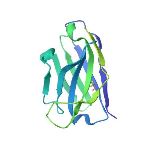
NA-22 fragment antigen binding heavy chainC [auth K],
F,
H [auth J],
L [auth H]
227Homo sapiensMutation(s): 0 
Entity Groups  
Sequence Clusters30% Identity50% Identity70% Identity90% Identity95% Identity100% Identity
Sequence Annotations
Expand
  • Reference Sequence
Oligosaccharides

Help

Entity ID: 4
MoleculeChains Length2D Diagram Glycosylation3D Interactions
2-acetamido-2-deoxy-beta-D-glucopyranose-(1-4)-2-acetamido-2-deoxy-beta-D-glucopyranose
M, O, Q, T
2N-Glycosylation
Glycosylation Resources
GlyTouCan:  G42666HT
GlyCosmos:  G42666HT
GlyGen:  G42666HT
Entity ID: 5
MoleculeChains Length2D Diagram Glycosylation3D Interactions
alpha-D-mannopyranose-(1-2)-alpha-D-mannopyranose-(1-2)-alpha-D-mannopyranose-(1-3)-[alpha-D-mannopyranose-(1-3)-[alpha-D-mannopyranose-(1-6)]alpha-D-mannopyranose-(1-6)]beta-D-mannopyranose-(1-4)-2-acetamido-2-deoxy-beta-D-glucopyranose-(1-4)-2-acetamido-2-deoxy-beta-D-glucopyranose
N, P, R, S
9N-Glycosylation
Glycosylation Resources
GlyTouCan:  G68668TB
GlyCosmos:  G68668TB
GlyGen:  G68668TB
Experimental Data & Validation

Experimental Data

  • Method: ELECTRON MICROSCOPY
  • Resolution: 3.00 Å
  • Aggregation State: PARTICLE 
  • Reconstruction Method: SINGLE PARTICLE 
EM Software:
TaskSoftware PackageVersion
MODEL REFINEMENTCoot
MODEL REFINEMENTRosetta3.10
RECONSTRUCTIONcryoSPARC2

Structure Validation

View Full Validation Report


Entry History & Funding Information

Deposition Data


Funding OrganizationLocationGrant Number
National Institutes of Health/National Institute Of Allergy and Infectious Diseases (NIH/NIAID)United StatesU19 AI117905
National Institutes of Health/National Institute Of Allergy and Infectious Diseases (NIH/NIAID)United StatesHHSN 272201400024C

Revision History  (Full details and data files)

  • Version 1.0: 2019-12-04
    Type: Initial release
  • Version 1.1: 2019-12-25
    Changes: Database references
  • Version 2.0: 2020-07-29
    Type: Remediation
    Reason: Carbohydrate remediation
    Changes: Atomic model, Data collection, Derived calculations, Structure summary
  • Version 2.1: 2023-04-05
    Changes: Database references, Refinement description
  • Version 2.2: 2024-10-09
    Changes: Data collection, Refinement description, Structure summary