7DF7

Crystal structure of human V-1 in the apo form


Experimental Data Snapshot

  • Method: X-RAY DIFFRACTION
  • Resolution: 2.30 Å
  • R-Value Free: 0.248 
  • R-Value Work: 0.218 
  • R-Value Observed: 0.220 

wwPDB Validation   3D Report Full Report


This is version 1.1 of the entry. See complete history


Literature

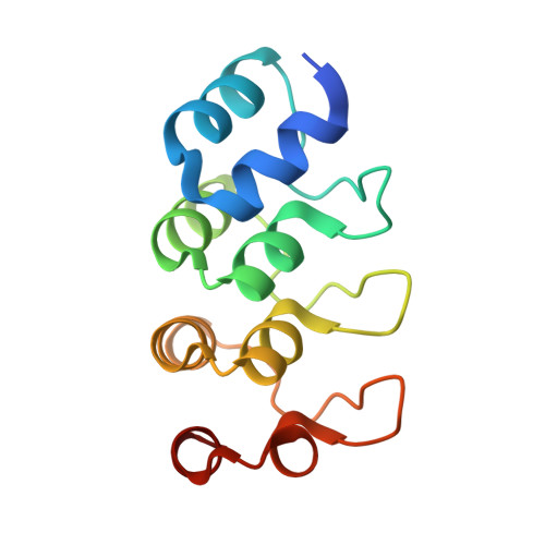
Crystal structure of human V-1 in the apo form.

Takeda, S.Koike, R.Nagae, T.Fujiwara, I.Narita, A.Maeda, Y.Ota, M.

(2021) Acta Crystallogr F Struct Biol Commun 77: 13-21

  • DOI: https://doi.org/10.1107/S2053230X20016829
  • Primary Citation of Related Structures:  
    7DF7

  • PubMed Abstract: 

    V-1, also known as myotrophin, is a 13 kDa ankyrin-repeat protein that binds and inhibits the heterodimeric actin capping protein (CP), which is a key regulator of cytoskeletal actin dynamics. The crystal structure of V-1 in complex with CP revealed that V-1 recognizes CP via residues spanning several ankyrin repeats. Here, the crystal structure of human V-1 is reported in the absence of the specific ligand at 2.3 Å resolution. In the asymmetric unit, the crystal contains two V-1 monomers that exhibit nearly identical structures (C α r.m.s.d. of 0.47 Å). The overall structures of the two apo V-1 chains are also highly similar to that of CP-bound V-1 (C α r.m.s.d.s of <0.50 Å), indicating that CP does not induce a large conformational change in V-1. Detailed structural comparisons using the computational program All Atom Motion Tree revealed that CP binding can be accomplished by minor side-chain rearrangements of several residues. These findings are consistent with the known biological role of V-1, in which it globally inhibits CP in the cytoplasm.


  • Organizational Affiliation

    Graduate School of Science, Nagoya University, Furo-cho, Chikusa-ku, Nagoya 464-8601, Japan.


Macromolecules
Find similar proteins by:  (by identity cutoff)  |  3D Structure
Entity ID: 1
MoleculeChains Sequence LengthOrganismDetailsImage
Myotrophin
A, B
123Homo sapiensMutation(s): 0 
Gene Names: MTPN
UniProt & NIH Common Fund Data Resources
Find proteins for P58546 (Homo sapiens)
Explore P58546 
Go to UniProtKB:  P58546
PHAROS:  P58546
GTEx:  ENSG00000105887 
Entity Groups  
Sequence Clusters30% Identity50% Identity70% Identity90% Identity95% Identity100% Identity
UniProt GroupP58546
Sequence Annotations
Expand
  • Reference Sequence
Experimental Data & Validation

Experimental Data

  • Method: X-RAY DIFFRACTION
  • Resolution: 2.30 Å
  • R-Value Free: 0.248 
  • R-Value Work: 0.218 
  • R-Value Observed: 0.220 
  • Space Group: P 21 21 21
Unit Cell:
Length ( Å )Angle ( ˚ )
a = 49.62α = 90
b = 56.28β = 90
c = 81.1γ = 90
Software Package:
Software NamePurpose
PHENIXrefinement
XDSdata reduction
XSCALEdata scaling
MOLREPphasing

Structure Validation

View Full Validation Report


Entry History & Funding Information

Deposition Data


Funding OrganizationLocationGrant Number
Japan Society for the Promotion of Science (JSPS)Japan16K17708
Japan Society for the Promotion of Science (JSPS)Japan17K07373
Japan Society for the Promotion of Science (JSPS)Japan20K06522

Revision History  (Full details and data files)

  • Version 1.0: 2021-01-20
    Type: Initial release
  • Version 1.1: 2023-11-29
    Changes: Data collection, Database references, Refinement description