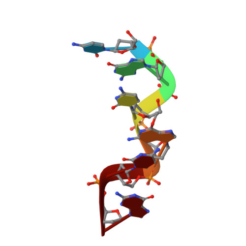
Crystal and molecular structure of a DNA duplex containing the carcinogenic lesion O6-methylguanine.
Ginell, S.L., Kuzmich, S., Jones, R.A., Berman, H.M.(1990) Biochemistry 29: 10461-10465
- PubMed: 2271657
- DOI: https://doi.org/10.1021/bi00498a005
- Primary Citation of Related Structures:
1D24 - PubMed Abstract:
The crystal and molecular structure of the first DNA duplex containing the carcinogenic lesion O6MeG has been determined to a resolution of 1.9 A and refined to an R factor of 19%. (d[CGC-(O6Me)GCG])2 crystallizes in the left-handed Z DNA form and has crystal parameters and conformational features similar to those of the parent sequence [d(CG)3]2. The methyl groups on O6 of G4 and G10 have C5-C6-O6-O6Me torsion angles of 73 degrees and 56 degrees, respectively, and protrude onto the major groove surface. The base-pairing conformation for the methylated G.C base pairs is of the Watson-Crick type as opposed to a wobble-type conformation that had been proposed in a B DNA fragment. As in other Z DNA structures, a spine of hydration is seen in the minor groove.
Organizational Affiliation:
Department of Chemistry, Wright-Rieman Laboratories, Rutgers University, Piscataway, New Jersey 08855-0939.