1ZGS

Parkia platycephala seed lectin in complex with 5-bromo-4-chloro-3-indolyl-a-D-mannose


Experimental Data Snapshot

  • Method: X-RAY DIFFRACTION
  • Resolution: 2.50 Å
  • R-Value Free: 
    0.244 (Depositor), 0.240 (DCC) 
  • R-Value Work: 
    0.208 (Depositor), 0.210 (DCC) 
  • R-Value Observed: 
    0.212 (Depositor) 

wwPDB Validation   3D Report Full Report


Ligand Structure Quality Assessment 

Created with Raphaël 2.3.0Worse 01 BetterLigand structure goodness of fit to experimental dataBest fitted XMMClick on this verticalbar to view details

This is version 1.4 of the entry. See complete history


Literature

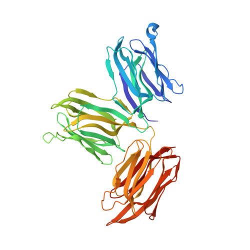
The first crystal structure of a mimosoideae lectin reveals a novel quaternary arrangement of a widespread domain.

Gallego Del Sol, F.Nagano, C.Cavada, B.S.Calvete, J.J.

(2005) J Mol Biol 353: 574-583

  • DOI: https://doi.org/10.1016/j.jmb.2005.08.055
  • Primary Citation of Related Structures:  
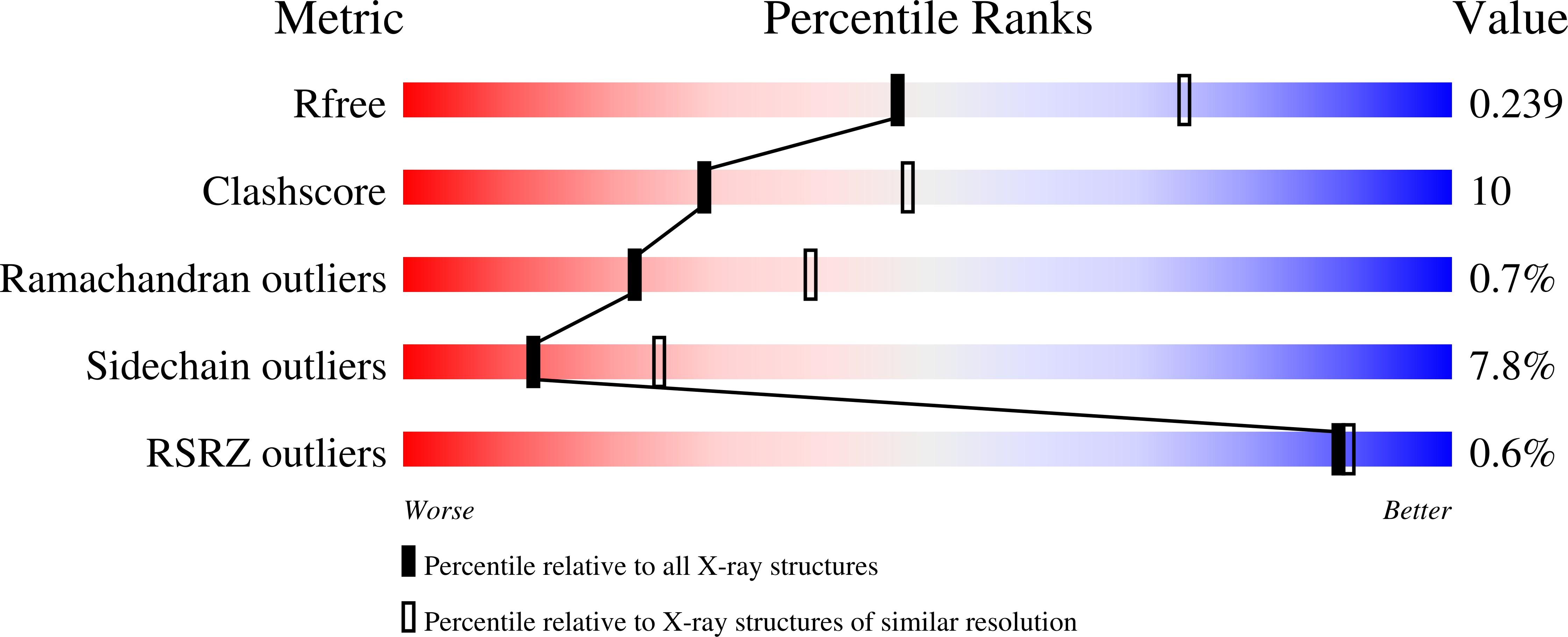
    1ZGR, 1ZGS

  • PubMed Abstract: 

    The crystal structures of the apo and mannose-bound Parkia platycephala seed lectin represent the first structure of a Mimosoideae lectin and a novel circular arrangement of beta-prism domains, and highlight the adaptability of the beta-prism fold as a building block in the evolution of plant lectins. The P.platycephala lectin is a dimer both in solution and in the crystals. Mannose binding to each of the three homologous carbohydrate-recognition domains of the lectin occurs through different modes, and restrains the flexibility of surface-exposed loops and residues involved in carbohydrate recognition. The planar array of carbohydrate-binding sites on the rim of the toroid-shaped structure of the P.platycephala lectin dimer immediately suggests a mechanism to promote multivalent interactions leading to cross-linking of carbohydrate ligands as part of the host strategy against phytopredators and pathogens. The cyclic structure of the P.platycephala lectin points to the convergent evolution of a structural principle for the construction of lectins involved in host defense or in attacking other organisms.


  • Organizational Affiliation

    Instituto de Biomedicina de Valencia, CSIC, E-46010 Valencia, Spain.


Macromolecules
Find similar proteins by:  (by identity cutoff)  |  3D Structure
Entity ID: 1
MoleculeChains Sequence LengthOrganismDetailsImage
Mannose/glucose-specific lectin
A, B
447Parkia platycephalaMutation(s): 0 
UniProt
Find proteins for P83304 (Parkia platycephala)
Explore P83304 
Go to UniProtKB:  P83304
Entity Groups  
Sequence Clusters30% Identity50% Identity70% Identity90% Identity95% Identity100% Identity
UniProt GroupP83304
Sequence Annotations
Expand
  • Reference Sequence
Experimental Data & Validation

Experimental Data

  • Method: X-RAY DIFFRACTION
  • Resolution: 2.50 Å
  • R-Value Free:  0.244 (Depositor), 0.240 (DCC) 
  • R-Value Work:  0.208 (Depositor), 0.210 (DCC) 
  • R-Value Observed: 0.212 (Depositor) 
Space Group: P 1 21 1
Unit Cell:
Length ( Å )Angle ( ˚ )
a = 80.239α = 90
b = 114.177β = 119.88
c = 80.304γ = 90
Software Package:
Software NamePurpose
REFMACrefinement
MOSFLMdata reduction
CCP4data scaling
SOLVEphasing

Structure Validation

View Full Validation Report


Ligand Structure Quality Assessment 

Created with Raphaël 2.3.0Worse 01 BetterLigand structure goodness of fit to experimental dataBest fitted XMMClick on this verticalbar to view details

Entry History 

Deposition Data

Revision History  (Full details and data files)

  • Version 1.0: 2005-10-11
    Type: Initial release
  • Version 1.1: 2008-04-30
    Changes: Version format compliance
  • Version 1.2: 2011-07-13
    Changes: Version format compliance
  • Version 1.3: 2020-07-29
    Type: Remediation
    Reason: Carbohydrate remediation
    Changes: Advisory, Derived calculations, Structure summary
  • Version 1.4: 2024-03-13
    Changes: Advisory, Data collection, Database references, Structure summary