3FYS

Crystal Structure of DegV, a fatty acid binding protein from Bacillus subtilis


Experimental Data Snapshot

  • Method: X-RAY DIFFRACTION
  • Resolution: 2.50 Å
  • R-Value Free: 
    0.225 (Depositor), 0.150 (DCC) 
  • R-Value Work: 
    0.148 (Depositor), 0.150 (DCC) 
  • R-Value Observed: 
    0.152 (Depositor) 

wwPDB Validation   3D Report Full Report


Ligand Structure Quality Assessment 

Created with Raphaël 2.3.0Worse 01 BetterLigand structure goodness of fit to experimental dataBest fitted PLMClick on this verticalbar to view details

This is version 1.2 of the entry. See complete history


Literature

Structure of a fatty acid-binding protein from Bacillus subtilis determined by sulfur-SAD phasing using in-house chromium radiation

Nan, J.Zhou, Y.F.Yang, C.Brostromer, E.Kristensen, O.Su, X.-D.

(2009) Acta Crystallogr D Biol Crystallogr 65: 440-448

  • DOI: https://doi.org/10.1107/S0907444909007756
  • Primary Citation of Related Structures:  
    3FYS

  • PubMed Abstract: 

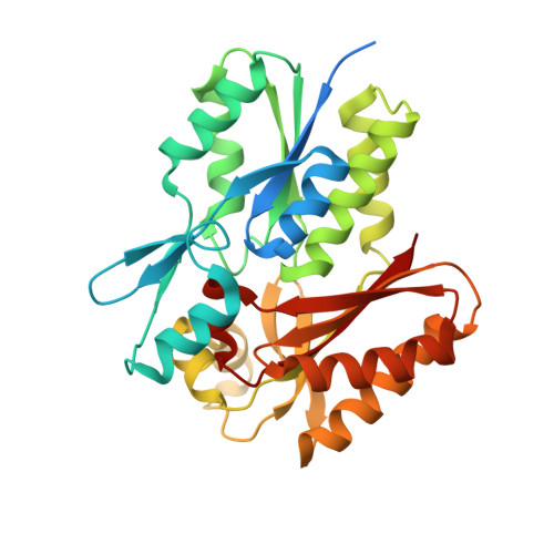
    Sulfur single-wavelength anomalous dispersion (S-SAD) and halide-soaking methods are increasingly being used for ab initio phasing. With the introduction of in-house Cr X-ray sources, these methods benefit from the enhanced anomalous scattering of S and halide atoms, respectively. Here, these methods were combined to determine the crystal structure of BsDegV, a DegV protein-family member from Bacillus subtilis. The protein was cocrystallized with bromide and low-redundancy data were collected to 2.5 A resolution using Cr Kalpha radiation. 17 heavy-atom sites (ten sulfurs and seven bromides) were located using standard methods. The anomalous scattering of some of the BsDegV S atoms and Br atoms was weak, thus neither sulfurs nor bromides could be used alone for structure determination using the collected data. When all 17 heavy-atom sites were used for SAD phasing, an easily interpretable electron-density map was obtained after density modification. The model of BsDegV was built automatically and a palmitate was found tightly bound in the active site. Sequence alignment and comparisons with other known DegV structures provided further insight into the specificity of fatty-acid selection and recognition within this protein family.


  • Organizational Affiliation

    National Laboratory of Protein Engineering and Plant Genetic Engineering, College of Life Sciences, Peking University, Beijing, People's Republic of China.


Macromolecules
Find similar proteins by:  (by identity cutoff)  |  3D Structure
Entity ID: 1
MoleculeChains Sequence LengthOrganismDetailsImage
Protein degV315Bacillus subtilisMutation(s): 0 
Gene Names: BSU35480degVyviA
UniProt
Find proteins for P32436 (Bacillus subtilis (strain 168))
Explore P32436 
Go to UniProtKB:  P32436
Entity Groups  
Sequence Clusters30% Identity50% Identity70% Identity90% Identity95% Identity100% Identity
UniProt GroupP32436
Sequence Annotations
Expand
  • Reference Sequence
Small Molecules
Ligands 3 Unique
IDChains Name / Formula / InChI Key2D Diagram3D Interactions
PLM
Query on PLM

Download Ideal Coordinates CCD File 
B [auth A]PALMITIC ACID
C16 H32 O2
IPCSVZSSVZVIGE-UHFFFAOYSA-N
BR
Query on BR

Download Ideal Coordinates CCD File 
D [auth A]
E [auth A]
F [auth A]
G [auth A]
H [auth A]
BROMIDE ION
Br
CPELXLSAUQHCOX-UHFFFAOYSA-M
EDO
Query on EDO

Download Ideal Coordinates CCD File 
C [auth A]1,2-ETHANEDIOL
C2 H6 O2
LYCAIKOWRPUZTN-UHFFFAOYSA-N
Experimental Data & Validation

Experimental Data

  • Method: X-RAY DIFFRACTION
  • Resolution: 2.50 Å
  • R-Value Free:  0.225 (Depositor), 0.150 (DCC) 
  • R-Value Work:  0.148 (Depositor), 0.150 (DCC) 
  • R-Value Observed: 0.152 (Depositor) 
Space Group: P 1 21 1
Unit Cell:
Length ( Å )Angle ( ˚ )
a = 42.1α = 90
b = 84.7β = 94.1
c = 42.2γ = 90
Software Package:
Software NamePurpose
HKL-2000data collection
SOLVEphasing
PHENIXrefinement
HKL-2000data reduction
HKL-2000data scaling

Structure Validation

View Full Validation Report


Ligand Structure Quality Assessment 

Created with Raphaël 2.3.0Worse 01 BetterLigand structure goodness of fit to experimental dataBest fitted PLMClick on this verticalbar to view details

Entry History 

Deposition Data

Revision History  (Full details and data files)

  • Version 1.0: 2009-05-12
    Type: Initial release
  • Version 1.1: 2011-07-13
    Changes: Version format compliance
  • Version 1.2: 2024-03-20
    Changes: Data collection, Database references, Derived calculations