3HNB

Factor VIII Trp2313-His2315 segment is involved in membrane binding as shown by crystal structure of complex between factor VIII C2 domain and an inhibitor


Experimental Data Snapshot

  • Method: X-RAY DIFFRACTION
  • Resolution: 1.15 Å
  • R-Value Free: 
    0.220 (Depositor), 0.220 (DCC) 
  • R-Value Work: 
    0.185 (Depositor), 0.180 (DCC) 
  • R-Value Observed: 
    0.187 (Depositor) 

Starting Model: experimental
View more details

wwPDB Validation   3D Report Full Report


Ligand Structure Quality Assessment 

Created with Raphaël 2.3.0Worse 01 BetterLigand structure goodness of fit to experimental dataBest fitted 768Click on this verticalbar to view details

This is version 1.3 of the entry. See complete history


Literature

Trp2313-His2315 of factor VIII C2 domain is involved in membrane binding: structure of a complex between the C2 domain and an inhibitor of membrane binding.

Liu, Z.Lin, L.Yuan, C.Nicolaes, G.A.Chen, L.Meehan, E.J.Furie, B.Furie, B.Huang, M.

(2010) J Biol Chem 285: 8824-8829

  • DOI: https://doi.org/10.1074/jbc.M109.080168
  • Primary Citation of Related Structures:  
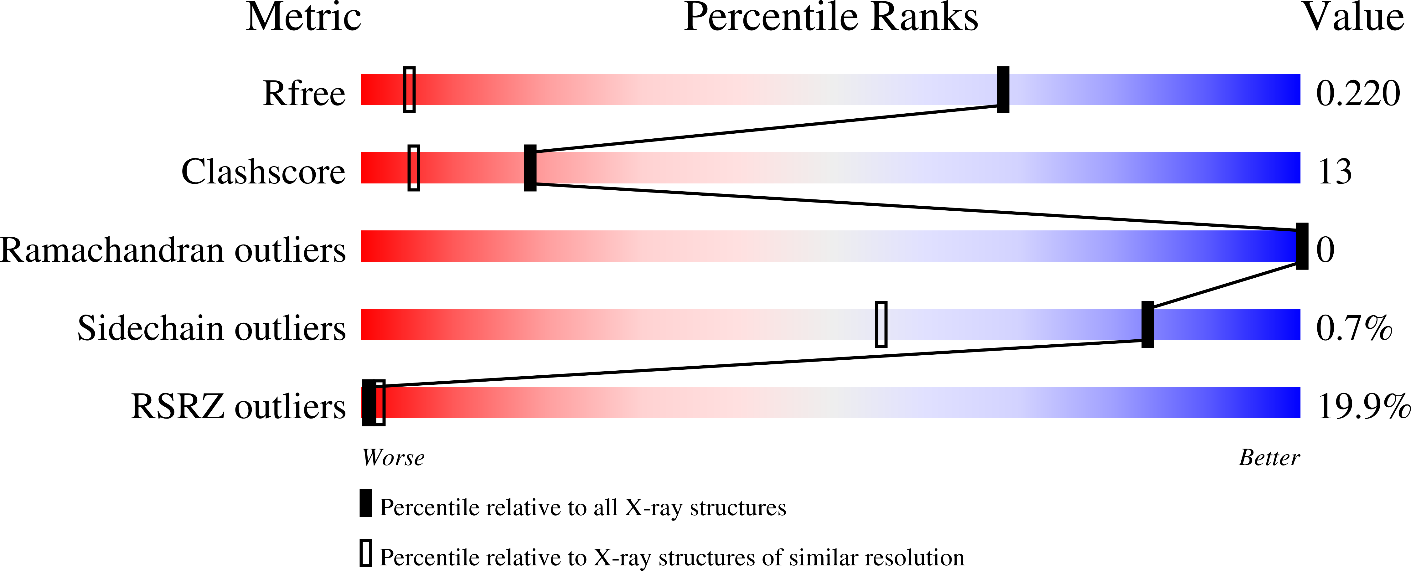
    3HNB, 3HNY, 3HOB

  • PubMed Abstract: 

    Factor VIII (FVIII) plays a critical role in blood coagulation by forming the tenase complex with factor IXa and calcium ions on a membrane surface containing negatively charged phospholipids. The tenase complex activates factor X during blood coagulation. The carboxyl-terminal C2 domain of FVIII is the main membrane-binding and von Willebrand factor-binding region of the protein. Mutations of FVIII cause hemophilia A, whereas elevation of FVIII activity is a risk factor for thromboembolic diseases. The C2 domain-membrane interaction has been proposed as a target of intervention for regulation of blood coagulation. A number of molecules that interrupt FVIII or factor V (FV) binding to cell membranes have been identified through high throughput screening or structure-based design. We report crystal structures of the FVIII C2 domain under three new crystallization conditions, and a high resolution (1.15 A) crystal structure of the FVIII C2 domain bound to a small molecular inhibitor. The latter structure shows that the inhibitor binds to the surface of an exposed beta-strand of the C2 domain, Trp(2313)-His(2315). This result indicates that the Trp(2313)-His(2315) segment is an important constituent of the membrane-binding motif and provides a model to understand the molecular mechanism of the C2 domain membrane interaction.


  • Organizational Affiliation

    State Key Laboratory of Structural Chemistry, Fujian Institute of Research on the Structure of Matter, Chinese Academy of Sciences, Fuzhou, Fujian 350002, China.


Macromolecules
Find similar proteins by:  (by identity cutoff)  |  3D Structure
Entity ID: 1
MoleculeChains Sequence LengthOrganismDetailsImage
Coagulation factor VIIIA [auth M]159Homo sapiensMutation(s): 0 
Gene Names: F8
UniProt & NIH Common Fund Data Resources
Find proteins for P00451 (Homo sapiens)
Explore P00451 
Go to UniProtKB:  P00451
PHAROS:  P00451
GTEx:  ENSG00000185010 
Entity Groups  
Sequence Clusters30% Identity50% Identity70% Identity90% Identity95% Identity100% Identity
UniProt GroupP00451
Sequence Annotations
Expand
  • Reference Sequence
Small Molecules
Ligands 1 Unique
IDChains Name / Formula / InChI Key2D Diagram3D Interactions
768
Query on 768

Download Ideal Coordinates CCD File 
B [auth M](2R)-1-(2,4-dichlorophenoxy)-3-[(2E)-2-imino-3-(2-piperidin-1-ylethyl)-2,3-dihydro-1H-benzimidazol-1-yl]propan-2-ol
C23 H28 Cl2 N4 O2
QMRFKZWWZBZQKJ-MVXIZWJUSA-N
Binding Affinity Annotations 
IDSourceBinding Affinity
768 BindingDB:  3HNB IC50: 7800 (nM) from 1 assay(s)
Experimental Data & Validation

Experimental Data

  • Method: X-RAY DIFFRACTION
  • Resolution: 1.15 Å
  • R-Value Free:  0.220 (Depositor), 0.220 (DCC) 
  • R-Value Work:  0.185 (Depositor), 0.180 (DCC) 
  • R-Value Observed: 0.187 (Depositor) 
Space Group: P 21 21 21
Unit Cell:
Length ( Å )Angle ( ˚ )
a = 42.054α = 90
b = 55.775β = 90
c = 67.971γ = 90
Software Package:
Software NamePurpose
SCALAdata scaling
REFMACrefinement
PDB_EXTRACTdata extraction
HKL-2000data collection
HKL-2000data reduction
HKL-2000data scaling
MOLREPphasing

Structure Validation

View Full Validation Report


Ligand Structure Quality Assessment 

Created with Raphaël 2.3.0Worse 01 BetterLigand structure goodness of fit to experimental dataBest fitted 768Click on this verticalbar to view details

Entry History 

Deposition Data

Revision History  (Full details and data files)

  • Version 1.0: 2010-01-19
    Type: Initial release
  • Version 1.1: 2011-07-13
    Changes: Version format compliance
  • Version 1.2: 2023-11-01
    Changes: Data collection, Database references, Derived calculations, Refinement description
  • Version 1.3: 2024-11-06
    Changes: Structure summary