


3IGS
Structure of the Salmonella enterica N-acetylmannosamine-6-phosphate 2-epimerase
- PDB DOI: https://doi.org/10.2210/pdb3IGS/pdb
- Classification: ISOMERASE
- Organism(s): Salmonella enterica subsp. enterica serovar Typhimurium
- Expression System: Escherichia coli
- Mutation(s): No
- Deposited: 2009-07-28 Released: 2009-08-04
Experimental Data Snapshot
- Method: X-RAY DIFFRACTION
- Resolution: 1.50 Å
- R-Value Free: 0.177 (Depositor), 0.180 (DCC)
- R-Value Work: 0.147 (Depositor), 0.150 (DCC)
- R-Value Observed: 0.148 (Depositor)

This is version 1.4 of the entry. See complete history.


Macromolecules
Find similar proteins by:
(by identity cutoff) | 3D Structure
Entity ID: 1 | |||||
|---|---|---|---|---|---|
| Molecule | Chains | Sequence Length | Organism | Details | Image |
| N-acetylmannosamine-6-phosphate 2-epimerase 2 | 232 | Salmonella enterica subsp. enterica serovar Typhimurium | Mutation(s): 0 Gene Names: nanE2, STM3337 EC: 5.1.3.9 | 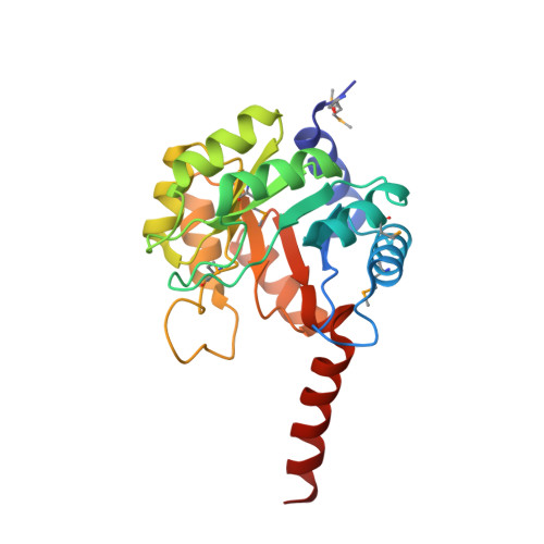 | |
UniProt | |||||
Find proteins for Q8ZLQ7 (Salmonella typhimurium (strain LT2 / SGSC1412 / ATCC 700720)) Explore Q8ZLQ7 Go to UniProtKB: Q8ZLQ7 | |||||
Entity Groups | |||||
| Sequence Clusters | 30% Identity50% Identity70% Identity90% Identity95% Identity100% Identity | ||||
| UniProt Group | Q8ZLQ7 | ||||
Sequence AnnotationsExpand | |||||
| |||||
Small Molecules
| Ligands 3 Unique | |||||
|---|---|---|---|---|---|
| ID | Chains | Name / Formula / InChI Key | 2D Diagram | 3D Interactions | |
| 16G Query on 16G | C [auth A] | 2-acetamido-2-deoxy-6-O-phosphono-alpha-D-glucopyranose C8 H16 N O9 P BRGMHAYQAZFZDJ-PVFLNQBWSA-N |  | ||
| SO4 Query on SO4 | D [auth A] E [auth A] F [auth A] G [auth A] H [auth A] | SULFATE ION O4 S QAOWNCQODCNURD-UHFFFAOYSA-L |  | ||
| CL Query on CL | K [auth A], S [auth B] | CHLORIDE ION Cl VEXZGXHMUGYJMC-UHFFFAOYSA-M |  | ||
| Modified Residues 1 Unique | |||||
|---|---|---|---|---|---|
| ID | Chains | Type | Formula | 2D Diagram | Parent |
| MSE Query on MSE | A, B | L-PEPTIDE LINKING | C5 H11 N O2 Se |  | MET |
Experimental Data & Validation
Experimental Data
- Method: X-RAY DIFFRACTION
- Resolution: 1.50 Å
- R-Value Free: 0.177 (Depositor), 0.180 (DCC)
- R-Value Work: 0.147 (Depositor), 0.150 (DCC)
- R-Value Observed: 0.148 (Depositor)
Diffraction Data: https://doi.org/10.18430/m33igs
Space Group: C 1 2 1
Unit Cell:
| Length ( Å ) | Angle ( ˚ ) |
|---|---|
| a = 183.29 | α = 90 |
| b = 63.32 | β = 91.28 |
| c = 41.8 | γ = 90 |
| Software Name | Purpose |
|---|---|
| DENZO | data reduction |
| SCALEPACK | data scaling |
| REFMAC | refinement |
| PDB_EXTRACT | data extraction |
| BLU-MAX | data collection |
| HKL-2000 | data reduction |
| SHARP | phasing |
Entry History
Deposition Data
- Released Date: 2009-08-04 Deposition Author(s): Anderson, S.M., Wawrzak, Z., Gordon, E., Skarina, T., Papazisi, L., Anderson, W.F., Savchenko, A., Center for Structural Genomics of Infectious Diseases (CSGID)
Revision History (Full details and data files)
- Version 1.0: 2009-08-04
Type: Initial release - Version 1.1: 2011-07-13
Changes: Version format compliance - Version 1.2: 2011-12-14
Changes: Structure summary - Version 1.3: 2017-11-01
Changes: Advisory, Refinement description - Version 1.4: 2020-07-29
Type: Remediation
Reason: Carbohydrate remediation
Changes: Data collection, Database references, Derived calculations, Structure summary

