3K8T

Structure of eukaryotic rnr large subunit R1 complexed with designed adp analog compound


Experimental Data Snapshot

  • Method: X-RAY DIFFRACTION
  • Resolution: 2.10 Å
  • R-Value Free: 
    0.290 (Depositor), 0.290 (DCC) 
  • R-Value Work: 
    0.252 (Depositor), 0.250 (DCC) 
  • R-Value Observed: 
    0.254 (Depositor) 

Starting Model: experimental
View more details

wwPDB Validation   3D Report Full Report


Ligand Structure Quality Assessment 

Created with Raphaël 2.3.0Worse 01 BetterLigand structure goodness of fit to experimental dataBest fitted DGTClick on this verticalbar to view detailsBest fitted 2A5Click on this verticalbar to view details

This is version 1.2 of the entry. See complete history


Literature

Structure-Based Design, Synthesis, and Evaluation of 2'-(2-Hydroxyethyl)-2'-deoxyadenosine and the 5'-Diphosphate Derivative as Ribonucleotide Reductase Inhibitors

Sun, D.Xu, H.Wijerathna, S.R.Dealwis, C.Lee, R.E.

(2009) ChemMedChem 4: 1649-1656

  • DOI: https://doi.org/10.1002/cmdc.200900236
  • Primary Citation of Related Structures:  
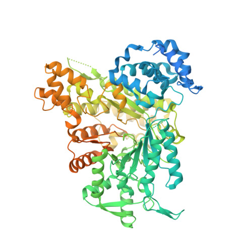
    3K8T

  • PubMed Abstract: 

    Analysis of the recently solved X-ray crystal structures of Saccharomyces cerevisiae ribonucleotide reductase I (ScRnr1) in complex with effectors and substrates led to the discovery of a conserved water molecule located at the active site that interacted with the 2'-hydroxy group of the nucleoside ribose. In this study 2'-(2-hydroxyethyl)-2'-deoxyadenosine 1 and the 5'-diphosphate derivative 2 were designed and synthesized to see if the conserved water molecule could be displaced by a hydroxymethylene group, to generate novel RNR inhibitors as potential antitumor agents. Herein we report the synthesis of analogues 1 and 2, and the co-crystal structure of adenosine diphosphate analogue 2 bound to ScRnr1, which shows the conserved water molecule is displaced as hypothesized.


  • Organizational Affiliation

    Department of Pharmaceutical Sciences, University of Tennessee Health Science Center, Memphis, TN 38163 (USA).


Macromolecules
Find similar proteins by:  (by identity cutoff)  |  3D Structure
Entity ID: 1
MoleculeChains Sequence LengthOrganismDetailsImage
Ribonucleoside-diphosphate reductase large chain 1888Saccharomyces cerevisiaeMutation(s): 0 
Gene Names: CRT7RIR1RNR1SDS12YER070W
EC: 1.17.4.1
UniProt
Find proteins for P21524 (Saccharomyces cerevisiae (strain ATCC 204508 / S288c))
Explore P21524 
Go to UniProtKB:  P21524
Entity Groups  
Sequence Clusters30% Identity50% Identity70% Identity90% Identity95% Identity100% Identity
UniProt GroupP21524
Sequence Annotations
Expand
  • Reference Sequence
Experimental Data & Validation

Experimental Data

  • Method: X-RAY DIFFRACTION
  • Resolution: 2.10 Å
  • R-Value Free:  0.290 (Depositor), 0.290 (DCC) 
  • R-Value Work:  0.252 (Depositor), 0.250 (DCC) 
  • R-Value Observed: 0.254 (Depositor) 
Space Group: P 21 21 2
Unit Cell:
Length ( Å )Angle ( ˚ )
a = 107.528α = 90
b = 116.915β = 90
c = 64.065γ = 90
Software Package:
Software NamePurpose
CCP4model building
REFMACrefinement
HKL-2000data reduction
HKL-2000data scaling
CCP4phasing

Structure Validation

View Full Validation Report


Ligand Structure Quality Assessment 

Created with Raphaël 2.3.0Worse 01 BetterLigand structure goodness of fit to experimental dataBest fitted DGTClick on this verticalbar to view detailsBest fitted 2A5Click on this verticalbar to view details

Entry History 

Deposition Data

Revision History  (Full details and data files)

  • Version 1.0: 2010-03-02
    Type: Initial release
  • Version 1.1: 2011-07-13
    Changes: Advisory, Version format compliance
  • Version 1.2: 2023-09-06
    Changes: Data collection, Database references, Derived calculations, Refinement description