Crystal structure of Putative sugar phosphate isomerase (AFE_0303) from Acidithiobacillus ferrooxidans ATCC 23270 at 1.85 A resolution
Joint Center for Structural Genomics (JCSG)To be published.
Experimental Data Snapshot
Entity ID: 1 | |||||
|---|---|---|---|---|---|
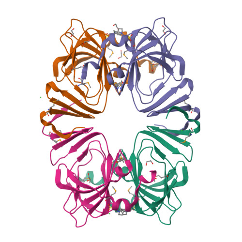
| Molecule | Chains | Sequence Length | Organism | Details | Image |
| Putative sugar phosphate isomerase | 162 | Acidithiobacillus ferrooxidans ATCC 23270 | Mutation(s): 0 Gene Names: AFE_0303, AFE_2805 |  | |
UniProt | |||||
Find proteins for B7J8Z8 (Acidithiobacillus ferrooxidans (strain ATCC 23270 / DSM 14882 / CIP 104768 / NCIMB 8455)) Explore B7J8Z8 Go to UniProtKB: B7J8Z8 | |||||
Entity Groups | |||||
| Sequence Clusters | 30% Identity50% Identity70% Identity90% Identity95% Identity100% Identity | ||||
| UniProt Group | B7J8Z8 | ||||
Sequence AnnotationsExpand | |||||
| |||||
| Ligands 3 Unique | |||||
|---|---|---|---|---|---|
| ID | Chains | Name / Formula / InChI Key | 2D Diagram | 3D Interactions | |
| CXS Query on CXS | G [auth A], L [auth B], T [auth C], W [auth D] | 3-CYCLOHEXYL-1-PROPYLSULFONIC ACID C9 H19 N O3 S PJWWRFATQTVXHA-UHFFFAOYSA-N |  | ||
| EDO Query on EDO | H [auth A] I [auth A] J [auth A] M [auth B] N [auth B] | 1,2-ETHANEDIOL C2 H6 O2 LYCAIKOWRPUZTN-UHFFFAOYSA-N |  | ||
| CL Query on CL | E [auth A], F [auth A], K [auth B] | CHLORIDE ION Cl VEXZGXHMUGYJMC-UHFFFAOYSA-M |  | ||
| Modified Residues 1 Unique | |||||
|---|---|---|---|---|---|
| ID | Chains | Type | Formula | 2D Diagram | Parent |
| MSE Query on MSE | A, B, C, D | L-PEPTIDE LINKING | C5 H11 N O2 Se |  | MET |
| Length ( Å ) | Angle ( ˚ ) |
|---|---|
| a = 75.876 | α = 90 |
| b = 93.744 | β = 90 |
| c = 139.042 | γ = 90 |
| Software Name | Purpose |
|---|---|
| REFMAC | refinement |
| PHENIX | refinement |
| SHELX | phasing |
| MolProbity | model building |
| XSCALE | data scaling |
| PDB_EXTRACT | data extraction |
| XDS | data reduction |
| SHELXD | phasing |
| autoSHARP | phasing |
RCSB PDB is hosted by

RCSB PDB is a member of the

All questions and comments will receive a response in a timely manner.