4DD8

ADAM-8 metalloproteinase domain with bound batimastat


Experimental Data Snapshot

  • Method: X-RAY DIFFRACTION
  • Resolution: 2.10 Å
  • R-Value Free: 
    0.255 (Depositor), 0.250 (DCC) 
  • R-Value Work: 
    0.188 (Depositor), 0.190 (DCC) 
  • R-Value Observed: 
    0.192 (Depositor) 

Starting Model: experimental
View more details

wwPDB Validation   3D Report Full Report


Ligand Structure Quality Assessment 

Created with Raphaël 2.3.0Worse 01 BetterLigand structure goodness of fit to experimental dataBest fitted BATClick on this verticalbar to view details

This is version 1.3 of the entry. See complete history


Literature

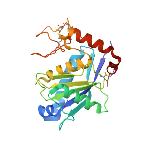
Structure of human ADAM-8 catalytic domain complexed with batimastat.

Hall, T.Shieh, H.S.Day, J.E.Caspers, N.Chrencik, J.E.Williams, J.M.Pegg, L.E.Pauley, A.M.Moon, A.F.Krahn, J.M.Fischer, D.H.Kiefer, J.R.Tomasselli, A.G.Zack, M.D.

(2012) Acta Crystallogr Sect F Struct Biol Cryst Commun 68: 616-621

  • DOI: https://doi.org/10.1107/S1744309112015618
  • Primary Citation of Related Structures:  
    4DD8

  • PubMed Abstract: 

    The role of ADAM-8 in cancer and inflammatory diseases such as allergy, arthritis and asthma makes it an attractive target for drug development. Therefore, the catalytic domain of human ADAM-8 was expressed, purified and crystallized in complex with a hydroxamic acid inhibitor, batimastat. The crystal structure of the enzyme-inhibitor complex was refined to 2.1 Å resolution. ADAM-8 has an overall fold similar to those of other ADAM members, including a central five-stranded β-sheet and a catalytic Zn(2+) ion. However, unique differences within the S1' binding loop of ADAM-8 are observed which might be exploited to confer specificity and selectivity to ADAM-8 competitive inhibitors for the treatment of diseases involving this enzyme.


  • Organizational Affiliation

    Pfizer Inc, 700 Chesterfield Parkway West, Chesterfield, MO 63017, USA.


Macromolecules
Find similar proteins by:  (by identity cutoff)  |  3D Structure
Entity ID: 1
MoleculeChains Sequence LengthOrganismDetailsImage
Disintegrin and metalloproteinase domain-containing protein 8
A, B, C, D
208Homo sapiensMutation(s): 0 
Gene Names: ADAM8MS2
EC: 3.4.24
UniProt & NIH Common Fund Data Resources
Find proteins for P78325 (Homo sapiens)
Explore P78325 
Go to UniProtKB:  P78325
PHAROS:  P78325
GTEx:  ENSG00000151651 
Entity Groups  
Sequence Clusters30% Identity50% Identity70% Identity90% Identity95% Identity100% Identity
UniProt GroupP78325
Sequence Annotations
Expand
  • Reference Sequence
Small Molecules
Ligands 6 Unique
IDChains Name / Formula / InChI Key2D Diagram3D Interactions
BAT
Query on BAT

Download Ideal Coordinates CCD File 
E [auth A],
M [auth B],
S [auth C],
Y [auth D]
4-(N-HYDROXYAMINO)-2R-ISOBUTYL-2S-(2-THIENYLTHIOMETHYL)SUCCINYL-L-PHENYLALANINE-N-METHYLAMIDE
C23 H31 N3 O4 S2
XFILPEOLDIKJHX-QYZOEREBSA-N
ZN
Query on ZN

Download Ideal Coordinates CCD File 
AA [auth D],
G [auth A],
O [auth B],
U [auth C]
ZINC ION
Zn
PTFCDOFLOPIGGS-UHFFFAOYSA-N
CA
Query on CA

Download Ideal Coordinates CCD File 
F [auth A],
N [auth B],
T [auth C],
Z [auth D]
CALCIUM ION
Ca
BHPQYMZQTOCNFJ-UHFFFAOYSA-N
K
Query on K

Download Ideal Coordinates CCD File 
K [auth A],
L [auth A]
POTASSIUM ION
K
NPYPAHLBTDXSSS-UHFFFAOYSA-N
CL
Query on CL

Download Ideal Coordinates CCD File 
CA [auth D],
I [auth A],
Q [auth B],
W [auth C]
CHLORIDE ION
Cl
VEXZGXHMUGYJMC-UHFFFAOYSA-M
NA
Query on NA

Download Ideal Coordinates CCD File 
BA [auth D]
DA [auth D]
H [auth A]
J [auth A]
P [auth B]
SODIUM ION
Na
FKNQFGJONOIPTF-UHFFFAOYSA-N
Experimental Data & Validation

Experimental Data

  • Method: X-RAY DIFFRACTION
  • Resolution: 2.10 Å
  • R-Value Free:  0.255 (Depositor), 0.250 (DCC) 
  • R-Value Work:  0.188 (Depositor), 0.190 (DCC) 
  • R-Value Observed: 0.192 (Depositor) 
Space Group: P 1 21 1
Unit Cell:
Length ( Å )Angle ( ˚ )
a = 91.6α = 90
b = 50.9β = 102.4
c = 93.5γ = 90
Software Package:
Software NamePurpose
ADSCdata collection
MOLREPphasing
PHENIXrefinement
REFMACrefinement
HKL-2000data reduction
HKL-2000data scaling

Structure Validation

View Full Validation Report


Ligand Structure Quality Assessment 

Created with Raphaël 2.3.0Worse 01 BetterLigand structure goodness of fit to experimental dataBest fitted BATClick on this verticalbar to view details

Entry History 

Revision History  (Full details and data files)

  • Version 1.0: 2012-06-06
    Type: Initial release
  • Version 1.1: 2012-06-27
    Changes: Database references
  • Version 1.2: 2023-09-13
    Changes: Data collection, Database references, Derived calculations, Refinement description
  • Version 1.3: 2024-11-06
    Changes: Structure summary