4LKV

Determinants of lipid substrate and membrane binding for the tetraacyldisaccharide-1-phosphate 4 -kinase LpxK


Experimental Data Snapshot

  • Method: X-RAY DIFFRACTION
  • Resolution: 3.51 Å
  • R-Value Free: 
    0.331 (Depositor), 0.330 (DCC) 
  • R-Value Work: 
    0.267 (Depositor), 0.270 (DCC) 
  • R-Value Observed: 
    0.273 (Depositor) 

wwPDB Validation   3D Report Full Report


Ligand Structure Quality Assessment 

Created with Raphaël 2.3.0Worse 01 BetterLigand structure goodness of fit to experimental dataBest fitted LP4Click on this verticalbar to view detailsBest fitted LP5Click on this verticalbar to view details

This is version 1.5 of the entry. See complete history


Literature

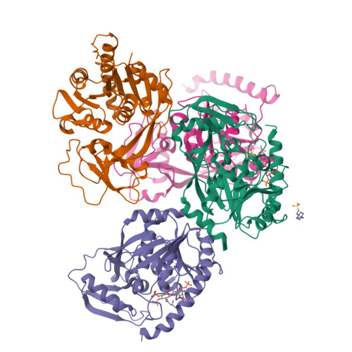
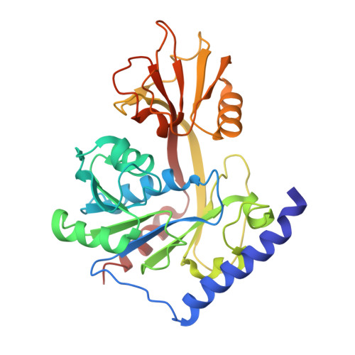
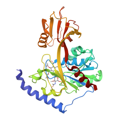
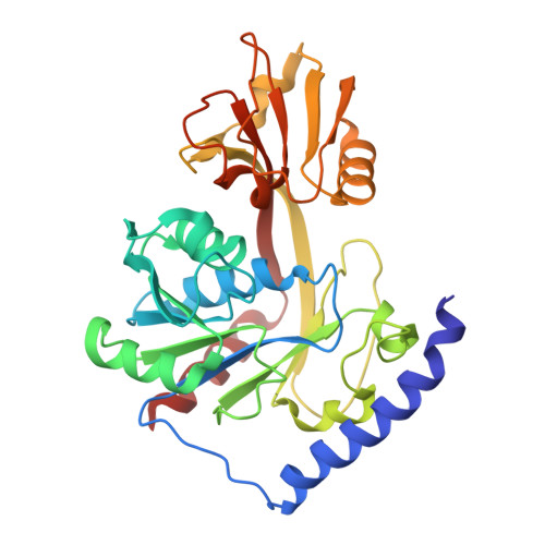
Structural Basis of Lipid Binding for the Membrane-embedded Tetraacyldisaccharide-1-phosphate 4'-Kinase LpxK.

Emptage, R.P.Tonthat, N.K.York, J.D.Schumacher, M.A.Zhou, P.

(2014) J Biol Chem 289: 24059-24068

  • DOI: https://doi.org/10.1074/jbc.M114.589986
  • Primary Citation of Related Structures:  
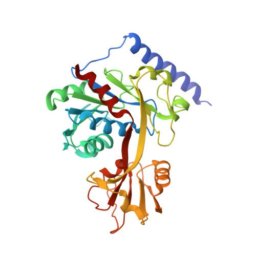
    4LKV

  • PubMed Abstract: 

    The membrane-bound tetraacyldisaccharide-1-phosphate 4'-kinase, LpxK, catalyzes the sixth step of the lipid A (Raetz) biosynthetic pathway and is a viable antibiotic target against emerging Gram-negative pathogens. We report the crystal structure of lipid IVA, the LpxK product, bound to the enzyme, providing a rare glimpse into interfacial catalysis and the surface scanning strategy by which many poorly understood lipid modification enzymes operate. Unlike the few previously structurally characterized proteins that bind lipid A or its precursors, LpxK binds almost exclusively to the glucosamine/phosphate moieties of the lipid molecule. Steady-state kinetic analysis of multiple point mutants of the lipid-binding pocket pinpoints critical residues involved in substrate binding, and characterization of N-terminal helix truncation mutants uncovers the role of this substructure as a hydrophobic membrane anchor. These studies make critical contributions to the limited knowledge surrounding membrane-bound enzymes that act upon lipid substrates and provide a structural template for designing small molecule inhibitors targeting this essential kinase.


  • Organizational Affiliation

    From the Department of Biochemistry, Duke University Medical Center, Durham, North Carolina 27710 and.


Macromolecules
Find similar proteins by:  (by identity cutoff)  |  3D Structure
Entity ID: 1
MoleculeChains Sequence LengthOrganismDetailsImage
Tetraacyldisaccharide 4'-kinase
A, B, C, D
315Aquifex aeolicus VF5Mutation(s): 0 
Gene Names: lpxKaq_1656
EC: 2.7.1.130
UniProt
Find proteins for O67572 (Aquifex aeolicus (strain VF5))
Explore O67572 
Go to UniProtKB:  O67572
Entity Groups  
Sequence Clusters30% Identity50% Identity70% Identity90% Identity95% Identity100% Identity
UniProt GroupO67572
Sequence Annotations
Expand
  • Reference Sequence
Small Molecules
Ligands 3 Unique
IDChains Name / Formula / InChI Key2D Diagram3D Interactions
LP4
Query on LP4

Download Ideal Coordinates CCD File 
F [auth A],
H [auth C]
2-deoxy-3-O-[(3R)-3-hydroxytetradecanoyl]-2-{[(3R)-3-hydroxytetradecanoyl]amino}-4-O-phosphono-beta-D-glucopyranose
C34 H66 N O12 P
AJRPJFBMHCTNGK-XQJZMFRCSA-N
LP5
Query on LP5

Download Ideal Coordinates CCD File 
G [auth A],
I [auth C]
(R)-((2R,3S,4R,5R,6R)-3-HYDROXY-2-(HYDROXYMETHYL)-5-((R)-3-HYDROXYTETRADECANAMIDO)-6-(PHOSPHONOOXY)TETRAHYDRO-2H-PYRAN-4-YL) 3-HYDROXYTETRADECANOATE
C34 H66 N O12 P
HEHQDWUWJVPREQ-XQJZMFRCSA-N
EPE
Query on EPE

Download Ideal Coordinates CCD File 
E [auth A]4-(2-HYDROXYETHYL)-1-PIPERAZINE ETHANESULFONIC ACID
C8 H18 N2 O4 S
JKMHFZQWWAIEOD-UHFFFAOYSA-N
Experimental Data & Validation

Experimental Data

  • Method: X-RAY DIFFRACTION
  • Resolution: 3.51 Å
  • R-Value Free:  0.331 (Depositor), 0.330 (DCC) 
  • R-Value Work:  0.267 (Depositor), 0.270 (DCC) 
  • R-Value Observed: 0.273 (Depositor) 
Space Group: C 2 2 21
Unit Cell:
Length ( Å )Angle ( ˚ )
a = 136.632α = 90
b = 140.691β = 90
c = 173.846γ = 90
Software Package:
Software NamePurpose
DENZOdata reduction
SCALEPACKdata scaling
PHASERphasing
RESOLVEphasing
PHENIXrefinement
PDB_EXTRACTdata extraction
SERGUIdata collection

Structure Validation

View Full Validation Report


Ligand Structure Quality Assessment 

Created with Raphaël 2.3.0Worse 01 BetterLigand structure goodness of fit to experimental dataBest fitted LP4Click on this verticalbar to view detailsBest fitted LP5Click on this verticalbar to view details

Entry History 

Deposition Data

Revision History  (Full details and data files)

  • Version 1.0: 2014-07-09
    Type: Initial release
  • Version 1.1: 2014-07-30
    Changes: Database references
  • Version 1.2: 2014-09-17
    Changes: Database references
  • Version 1.3: 2014-09-24
    Changes: Database references
  • Version 1.4: 2017-10-25
    Changes: Author supporting evidence, Refinement description
  • Version 1.5: 2024-02-28
    Changes: Data collection, Database references, Derived calculations, Refinement description