5GT9

The X-ray structure of 7beta-hydroxysteroid dehydrogenase


Experimental Data Snapshot

  • Method: X-RAY DIFFRACTION
  • Resolution: 1.70 Å
  • R-Value Free: 
    0.201 (Depositor), 0.210 (DCC) 
  • R-Value Work: 
    0.165 (Depositor), 0.180 (DCC) 
  • R-Value Observed: 
    0.167 (Depositor) 

Starting Model: experimental
View more details

wwPDB Validation   3D Report Full Report


Ligand Structure Quality Assessment 

Created with Raphaël 2.3.0Worse 01 BetterLigand structure goodness of fit to experimental dataBest fitted NAPClick on this verticalbar to view details

This is version 1.1 of the entry. See complete history


Literature

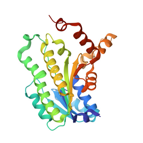
Structure of NADP(+)-bound 7 beta-hydroxysteroid dehydrogenase reveals two cofactor-binding modes

Wang, R.Wu, J.Jin, D.K.Chen, Y.Lv, Z.Chen, Q.Miao, Q.Huo, X.Wang, F.

(2017) Acta Crystallogr F Struct Biol Commun 73: 246-252

  • DOI: https://doi.org/10.1107/S2053230X17004460
  • Primary Citation of Related Structures:  
    5GT9

  • PubMed Abstract: 

    In mammals, bile acids/salts and their glycine and taurine conjugates are effectively recycled through enterohepatic circulation. 7β-Hydroxysteroid dehydrogenases (7β-HSDHs; EC 1.1.1.201), including that from the intestinal microbe Collinsella aerofaciens, catalyse the NADPH-dependent reversible oxidation of secondary bile-acid products to avoid potential toxicity. Here, the first structure of NADP + bound to dimeric 7β-HSDH is presented. In one active site, NADP + adopts a conventional binding mode similar to that displayed in related enzyme structures. However, in the other active site a unique binding mode is observed in which the orientation of the nicotinamide is different. Since 7β-HSDH has become an attractive target owing to the wide and important pharmaceutical use of its product ursodeoxycholic acid, this work provides a more detailed template to support rational protein engineering to improve the enzymatic activities of this useful biocatalyst, further improving the yield of ursodeoxycholic acid and its other applications.


  • Organizational Affiliation

    Wuxi Biortus Biosciences Co. Ltd, A5, 6 Dongsheng West Road, 214437 Jiangyin, Jiangsu, People's Republic of China.


Macromolecules
Find similar proteins by:  (by identity cutoff)  |  3D Structure
Entity ID: 1
MoleculeChains Sequence LengthOrganismDetailsImage
Oxidoreductase, short chain dehydrogenase/reductase family protein
A, B
263Collinsella aerofaciens ATCC 25986Mutation(s): 0 
Gene Names: COLAER_02088
EC: 1.1.1.201
UniProt
Find proteins for A4ECA9 (Collinsella aerofaciens (strain ATCC 25986 / DSM 3979 / JCM 10188 / KCTC 3647 / NCTC 11838 / VPI 1003))
Explore A4ECA9 
Go to UniProtKB:  A4ECA9
Entity Groups  
Sequence Clusters30% Identity50% Identity70% Identity90% Identity95% Identity100% Identity
UniProt GroupA4ECA9
Sequence Annotations
Expand
  • Reference Sequence
Experimental Data & Validation

Experimental Data

  • Method: X-RAY DIFFRACTION
  • Resolution: 1.70 Å
  • R-Value Free:  0.201 (Depositor), 0.210 (DCC) 
  • R-Value Work:  0.165 (Depositor), 0.180 (DCC) 
  • R-Value Observed: 0.167 (Depositor) 
Space Group: P 41 21 2
Unit Cell:
Length ( Å )Angle ( ˚ )
a = 72.73α = 90
b = 72.73β = 90
c = 171.31γ = 90
Software Package:
Software NamePurpose
REFMACrefinement
iMOSFLMdata processing

Structure Validation

View Full Validation Report


Ligand Structure Quality Assessment 

Created with Raphaël 2.3.0Worse 01 BetterLigand structure goodness of fit to experimental dataBest fitted NAPClick on this verticalbar to view details

Entry History & Funding Information

Deposition Data


Funding OrganizationLocationGrant Number
Jiangsu Innovation fund for Technology-based FirmsChinaBC2013062
Torch High Technology Industry Development Center, Ministry of Science and Technology, ChinaChina2015GH581522

Revision History  (Full details and data files)

  • Version 1.0: 2017-05-17
    Type: Initial release
  • Version 1.1: 2023-11-08
    Changes: Data collection, Database references, Refinement description