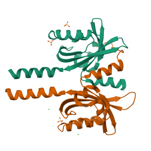
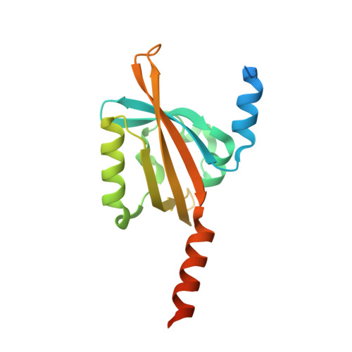
Structure of a LOV protein in apo-state and implications for construction of LOV-based optical tools.
Arinkin, V., Granzin, J., Rollen, K., Krauss, U., Jaeger, K.E., Willbold, D., Batra-Safferling, R.(2017) Sci Rep 7: 42971-42971
- PubMed: 28211532
- DOI: https://doi.org/10.1038/srep42971
- Primary Citation of Related Structures:
5LUV - PubMed Abstract:
Unique features of Light-Oxygen-Voltage (LOV) proteins like relatively small size (~12-19 kDa), inherent modularity, highly-tunable photocycle and oxygen-independent fluorescence have lately been exploited for the generation of optical tools. Structures of LOV domains reported so far contain a flavin chromophore per protein molecule. Here we report two new findings on the short LOV protein W619_1-LOV from Pseudomonas putida. First, the apo-state crystal structure of W619_1-LOV at 2.5 Å resolution reveals conformational rearrangements in the secondary structure elements lining the chromophore pocket including elongation of the Fα helix, shortening of the Eα-Fα loop and partial unfolding of the Eα helix. Second, the apo W619_1-LOV protein binds both natural and structurally modified flavin chromophores. Remarkably different photophysical and photochemical properties of W619_1-LOV bound to 7-methyl-8-chloro-riboflavin (8-Cl-RF) and lumichrome imply application of these variants as novel optical tools as they offer advantages such as no adduct state formation, and a broader choice of wavelengths for in vitro studies.
Organizational Affiliation:
Institute of Complex Systems, ICS-6: Structural Biochemistry, Forschungszentrum Jülich, 52425 Jülich, Germany.