5MJI

Crystal Structure of RosB with bound intermediate OHC-RP (8-demethyl-8-formylriboflavin-5'-phosphate)


Experimental Data Snapshot

  • Method: X-RAY DIFFRACTION
  • Resolution: 2.00 Å
  • R-Value Free: 
    0.209 (Depositor), 0.210 (DCC) 
  • R-Value Work: 
    0.186 (Depositor), 0.190 (DCC) 
  • R-Value Observed: 
    0.187 (Depositor) 

wwPDB Validation   3D Report Full Report


Ligand Structure Quality Assessment 

Created with Raphaël 2.3.0Worse 01 BetterLigand structure goodness of fit to experimental dataBest fitted 7O6Click on this verticalbar to view details

This is version 1.2 of the entry. See complete history


Literature

The Crystal Structure of RosB: Insights into the Reaction Mechanism of the First Member of a Family of Flavodoxin-like Enzymes.

Konjik, V.Brunle, S.Demmer, U.Vanselow, A.Sandhoff, R.Ermler, U.Mack, M.

(2017) Angew Chem Int Ed Engl 56: 1146-1151

  • DOI: https://doi.org/10.1002/anie.201610292
  • Primary Citation of Related Structures:  
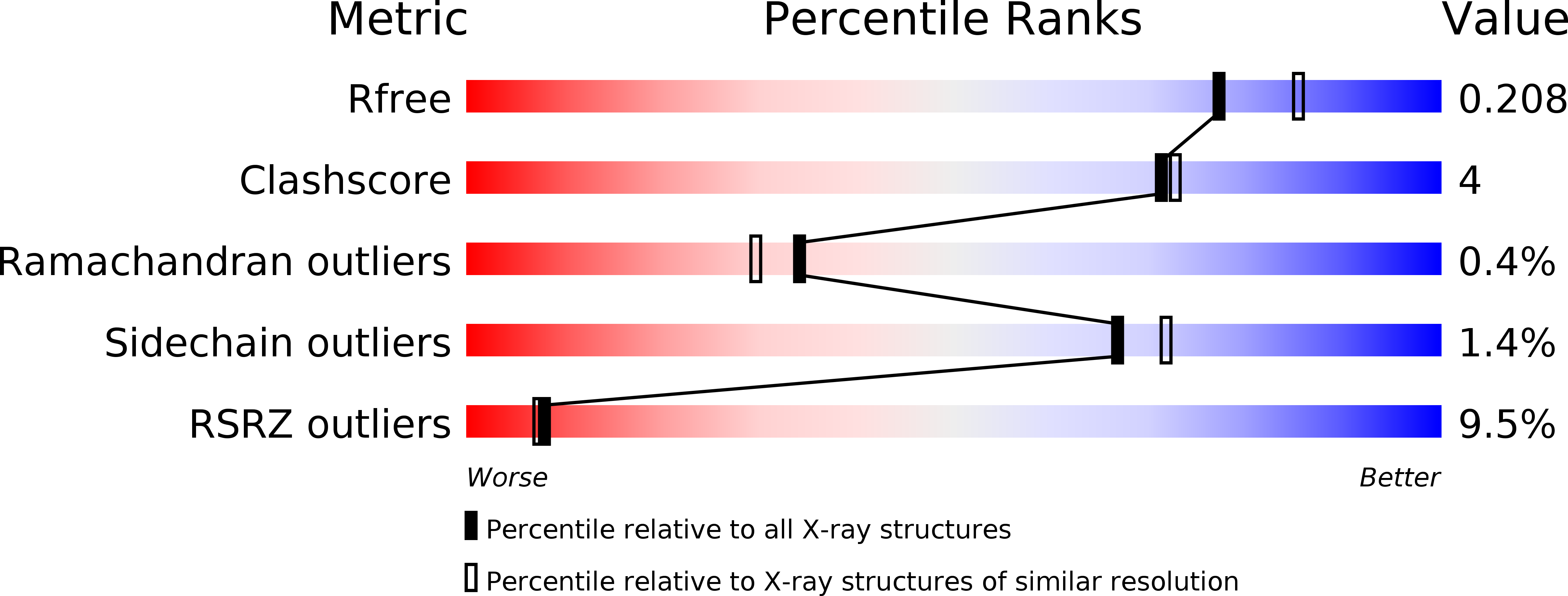
    5MJI

  • PubMed Abstract: 

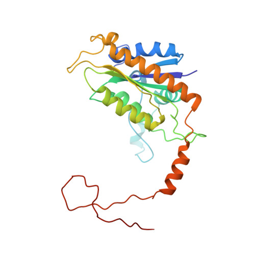
    8-demethyl-8-aminoriboflavin-5'-phosphate (AFP) synthase (RosB) catalyzes the key reaction of roseoflavin biosynthesis by forming AFP from riboflavin-5'-phosphate (RP) and glutamate via the intermediates 8-demethyl-8-formylriboflavin-5'-phosphate (OHC-RP) and 8-demethyl-8-carboxylriboflavin-5'-phosphate (HO 2 C-RP). To understand this reaction in which a methyl substituent of an aromatic ring is replaced by an amine we structurally characterized RosB in complex with OHC-RP (2.0 Å) and AFP (1.7 Å). RosB is composed of four flavodoxin-like subunits which have been upgraded with specific extensions and a unique C-terminal arm. It appears that RosB has evolved from an electron- or hydride-transferring flavoprotein to a sophisticated multi-step enzyme which uses RP as a substrate (and not as a cofactor). Structure-based active site analysis was complemented by mutational and isotope-based mass-spectrometric data to propose an enzymatic mechanism on an atomic basis.


  • Organizational Affiliation

    Mannheim University of Applied Sciences, Paul-Wittsack-Strasse 10, 68163, Mannheim, Germany.


Macromolecules
Find similar proteins by:  (by identity cutoff)  |  3D Structure
Entity ID: 1
MoleculeChains Sequence LengthOrganismDetailsImage
BRAMP domain protein257Streptomyces davaonensis JCM 4913Mutation(s): 0 
Gene Names: BN159_7989
EC: 2.6.1.114
UniProt
Find proteins for K4REZ6 (Streptomyces davaonensis (strain DSM 101723 / JCM 4913 / KCC S-0913 / 768))
Explore K4REZ6 
Go to UniProtKB:  K4REZ6
Entity Groups  
Sequence Clusters30% Identity50% Identity70% Identity90% Identity95% Identity100% Identity
UniProt GroupK4REZ6
Sequence Annotations
Expand
  • Reference Sequence
Small Molecules
Ligands 1 Unique
IDChains Name / Formula / InChI Key2D Diagram3D Interactions
7O6
Query on 7O6

Download Ideal Coordinates CCD File 
B [auth A][(2~{S},3~{R},4~{R})-5-[8-methanoyl-7-methyl-2,4-bis(oxidanylidene)-1~{H}-benzo[g]pteridin-10-ium-10-yl]-2,3,4-tris(oxidanyl)pentyl] dihydrogen phosphate
C17 H20 N4 O10 P
TZAPMZFMEMVUSE-MBNYWOFBSA-O
Experimental Data & Validation

Experimental Data

  • Method: X-RAY DIFFRACTION
  • Resolution: 2.00 Å
  • R-Value Free:  0.209 (Depositor), 0.210 (DCC) 
  • R-Value Work:  0.186 (Depositor), 0.190 (DCC) 
  • R-Value Observed: 0.187 (Depositor) 
Space Group: I 41 2 2
Unit Cell:
Length ( Å )Angle ( ˚ )
a = 108.47α = 90
b = 108.47β = 90
c = 178.22γ = 90
Software Package:
Software NamePurpose
PHENIXrefinement
XDSdata reduction
XSCALEdata scaling
PHENIXphasing

Structure Validation

View Full Validation Report


Ligand Structure Quality Assessment 

Created with Raphaël 2.3.0Worse 01 BetterLigand structure goodness of fit to experimental dataBest fitted 7O6Click on this verticalbar to view details

Entry History 

Deposition Data

Revision History  (Full details and data files)

  • Version 1.0: 2016-12-28
    Type: Initial release
  • Version 1.1: 2017-01-25
    Changes: Database references
  • Version 1.2: 2024-05-08
    Changes: Data collection, Database references