5UPH

Lipids bound lysosomal integral membrane protein 2


Experimental Data Snapshot

  • Method: X-RAY DIFFRACTION
  • Resolution: 3.00 Å
  • R-Value Free: 
    0.228 (Depositor), 0.240 (DCC) 
  • R-Value Work: 
    0.193 (Depositor), 0.210 (DCC) 
  • R-Value Observed: 
    0.195 (Depositor) 

Starting Model: experimental
View more details

wwPDB Validation   3D Report Full Report


Ligand Structure Quality Assessment 

Created with Raphaël 2.3.0Worse 01 BetterLigand structure goodness of fit to experimental dataBest fitted PCWClick on this verticalbar to view detailsBest fitted CLRClick on this verticalbar to view details

This is version 2.2 of the entry. See complete history


Literature

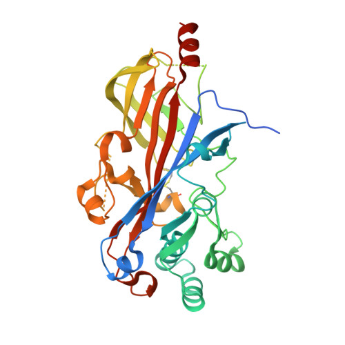
Lysosomal integral membrane protein-2 as a phospholipid receptor revealed by biophysical and cellular studies.

Conrad, K.S.Cheng, T.W.Ysselstein, D.Heybrock, S.Hoth, L.R.Chrunyk, B.A.Am Ende, C.W.Krainc, D.Schwake, M.Saftig, P.Liu, S.Qiu, X.Ehlers, M.D.

(2017) Nat Commun 8: 1908-1908

  • DOI: https://doi.org/10.1038/s41467-017-02044-8
  • Primary Citation of Related Structures:  
    5UPH

  • PubMed Abstract: 

    Lysosomal integral membrane protein-2 (LIMP-2/SCARB2) contributes to endosomal and lysosomal function. LIMP-2 deficiency is associated with neurological abnormalities and kidney failure and, as an acid glucocerebrosidase receptor, impacts Gaucher and Parkinson's diseases. Here we report a crystal structure of a LIMP-2 luminal domain dimer with bound cholesterol and phosphatidylcholine. Binding of these lipids alters LIMP-2 from functioning as a glucocerebrosidase-binding monomer toward a dimeric state that preferentially binds anionic phosphatidylserine over neutral phosphatidylcholine. In cellular uptake experiments, LIMP-2 facilitates transport of phospholipids into murine fibroblasts, with a strong substrate preference for phosphatidylserine. Taken together, these biophysical and cellular studies define the structural basis and functional importance of a form of LIMP-2 for lipid trafficking. We propose a model whereby switching between monomeric and dimeric forms allows LIMP-2 to engage distinct binding partners, a mechanism that may be shared by SR-BI and CD36, scavenger receptor proteins highly homologous to LIMP-2.


  • Organizational Affiliation

    Medicinal Sciences, Pfizer Worldwide R&D, Eastern Point Road, Groton, CT, 06340, USA.


Macromolecules
Find similar proteins by:  (by identity cutoff)  |  3D Structure
Entity ID: 1
MoleculeChains Sequence LengthOrganismDetailsImage
Lysosome membrane protein 2
A, B
410Homo sapiensMutation(s): 0 
Gene Names: SCARB2CD36L2LIMP2LIMPII
Membrane Entity: Yes 
UniProt & NIH Common Fund Data Resources
Find proteins for Q14108 (Homo sapiens)
Explore Q14108 
Go to UniProtKB:  Q14108
PHAROS:  Q14108
GTEx:  ENSG00000138760 
Entity Groups  
Sequence Clusters30% Identity50% Identity70% Identity90% Identity95% Identity100% Identity
UniProt GroupQ14108
Glycosylation
Glycosylation Sites: 4Go to GlyGen: Q14108-1
Sequence Annotations
Expand
  • Reference Sequence
Oligosaccharides

Help

Entity ID: 2
MoleculeChains Length2D Diagram Glycosylation3D Interactions
alpha-D-mannopyranose-(1-3)-beta-D-mannopyranose-(1-4)-2-acetamido-2-deoxy-beta-D-glucopyranose-(1-4)-2-acetamido-2-deoxy-beta-D-glucopyranose
C, I
4N-Glycosylation
Glycosylation Resources
GlyTouCan:  G81315DD
GlyCosmos:  G81315DD
GlyGen:  G81315DD
Entity ID: 3
MoleculeChains Length2D Diagram Glycosylation3D Interactions
2-acetamido-2-deoxy-beta-D-glucopyranose-(1-4)-2-acetamido-2-deoxy-beta-D-glucopyranose
D, H
2N-Glycosylation
Glycosylation Resources
GlyTouCan:  G42666HT
GlyCosmos:  G42666HT
GlyGen:  G42666HT
Entity ID: 4
MoleculeChains Length2D Diagram Glycosylation3D Interactions
6-O-phosphono-beta-D-mannopyranose-(1-2)-alpha-D-mannopyranose-(1-6)-alpha-D-mannopyranose-(1-6)-[alpha-D-mannopyranose-(1-3)]beta-D-mannopyranose-(1-4)-2-acetamido-2-deoxy-beta-D-glucopyranose-(1-4)-2-acetamido-2-deoxy-beta-D-glucopyranose
E
7N-Glycosylation
Glycosylation Resources
GlyTouCan:  G83390WW
GlyCosmos:  G83390WW
Entity ID: 5
MoleculeChains Length2D Diagram Glycosylation3D Interactions
alpha-D-mannopyranose-(1-3)-[alpha-D-mannopyranose-(1-6)]beta-D-mannopyranose-(1-4)-2-acetamido-2-deoxy-beta-D-glucopyranose-(1-4)-2-acetamido-2-deoxy-beta-D-glucopyranose
F, G, J
5N-Glycosylation
Glycosylation Resources
GlyTouCan:  G22768VO
GlyCosmos:  G22768VO
GlyGen:  G22768VO
Experimental Data & Validation

Experimental Data

  • Method: X-RAY DIFFRACTION
  • Resolution: 3.00 Å
  • R-Value Free:  0.228 (Depositor), 0.240 (DCC) 
  • R-Value Work:  0.193 (Depositor), 0.210 (DCC) 
  • R-Value Observed: 0.195 (Depositor) 
Space Group: P 42 21 2
Unit Cell:
Length ( Å )Angle ( ˚ )
a = 139.04α = 90
b = 139.04β = 90
c = 178.28γ = 90
Software Package:
Software NamePurpose
BUSTERrefinement
XDSdata reduction
Aimlessdata scaling
PHASERphasing

Structure Validation

View Full Validation Report


Ligand Structure Quality Assessment 

Created with Raphaël 2.3.0Worse 01 BetterLigand structure goodness of fit to experimental dataBest fitted PCWClick on this verticalbar to view detailsBest fitted CLRClick on this verticalbar to view details

Entry History 

Deposition Data

Revision History  (Full details and data files)

  • Version 1.0: 2017-12-13
    Type: Initial release
  • Version 1.1: 2018-12-26
    Changes: Data collection, Database references
  • Version 2.0: 2020-07-29
    Type: Remediation
    Reason: Carbohydrate remediation
    Changes: Advisory, Atomic model, Data collection, Derived calculations, Structure summary
  • Version 2.1: 2023-10-04
    Changes: Data collection, Database references, Derived calculations, Refinement description, Structure summary
  • Version 2.2: 2024-11-06
    Changes: Structure summary