5XBI

The structure of BrlR-C domain bound to 3-amino-2-phenazino(a pyocyanin analog)


Experimental Data Snapshot

  • Method: X-RAY DIFFRACTION
  • Resolution: 1.40 Å
  • R-Value Free: 
    0.182 (Depositor), 0.180 (DCC) 
  • R-Value Work: 
    0.144 (Depositor), 0.140 (DCC) 
  • R-Value Observed: 
    0.146 (Depositor) 

wwPDB Validation   3D Report Full Report


Ligand Structure Quality Assessment 

Created with Raphaël 2.3.0Worse 01 BetterLigand structure goodness of fit to experimental dataBest fitted 81UClick on this verticalbar to view details

This is version 1.2 of the entry. See complete history


Literature

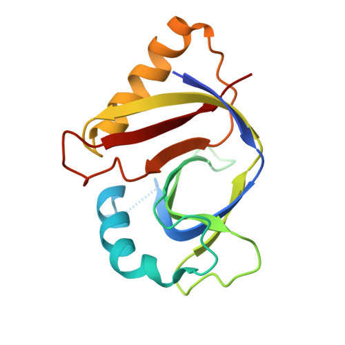
BrlR from Pseudomonas aeruginosa is a receptor for both cyclic di-GMP and pyocyanin.

Wang, F.He, Q.Yin, J.Xu, S.Hu, W.Gu, L.

(2018) Nat Commun 9: 2563-2563

  • DOI: https://doi.org/10.1038/s41467-018-05004-y
  • Primary Citation of Related Structures:  
    5XBI, 5XBT, 5XBW

  • PubMed Abstract: 

    The virulence factor pyocyanin and the intracellular second messenger cyclic diguanylate monophosphate (c-di-GMP) play key roles in regulating biofilm formation and multi-drug efflux pump expression in Pseudomonas aeruginosa. However, the crosstalk between these two signaling pathways remains unclear. Here we show that BrlR (PA4878), previously identified as a c-di-GMP responsive transcriptional regulator, acts also as a receptor for pyocyanin. Crystal structures of free BrlR and c-di-GMP-bound BrlR reveal that the DNA-binding domain of BrlR contains two separate c-di-GMP binding sites, both of which are involved in promoting brlR expression. In addition, we identify a pyocyanin-binding site on the C-terminal multidrug-binding domain based on the structure of the BrlR-C domain in complex with a pyocyanin analog. Biochemical analysis indicates that pyocyanin enhances BrlR-DNA binding and brlR expression in a concentration-dependent manner.


  • Organizational Affiliation

    State Key Laboratory of Microbial Technology, Shandong University, Jinan, 250100, Shandong, China.


Macromolecules
Find similar proteins by:  (by identity cutoff)  |  3D Structure
Entity ID: 1
MoleculeChains Sequence LengthOrganismDetailsImage
Probable transcriptional regulator
A, B
153Pseudomonas aeruginosa PAO1Mutation(s): 0 
Gene Names: PA4878
UniProt
Find proteins for Q9HUT5 (Pseudomonas aeruginosa (strain ATCC 15692 / DSM 22644 / CIP 104116 / JCM 14847 / LMG 12228 / 1C / PRS 101 / PAO1))
Explore Q9HUT5 
Go to UniProtKB:  Q9HUT5
Entity Groups  
Sequence Clusters30% Identity50% Identity70% Identity90% Identity95% Identity100% Identity
UniProt GroupQ9HUT5
Sequence Annotations
Expand
  • Reference Sequence
Experimental Data & Validation

Experimental Data

  • Method: X-RAY DIFFRACTION
  • Resolution: 1.40 Å
  • R-Value Free:  0.182 (Depositor), 0.180 (DCC) 
  • R-Value Work:  0.144 (Depositor), 0.140 (DCC) 
  • R-Value Observed: 0.146 (Depositor) 
Space Group: P 1 21 1
Unit Cell:
Length ( Å )Angle ( ˚ )
a = 39.705α = 90
b = 103.628β = 118.99
c = 39.774γ = 90
Software Package:
Software NamePurpose
PHENIXrefinement
HKL-3000data reduction
HKL-3000data scaling
PHENIXphasing

Structure Validation

View Full Validation Report


Ligand Structure Quality Assessment 

Created with Raphaël 2.3.0Worse 01 BetterLigand structure goodness of fit to experimental dataBest fitted 81UClick on this verticalbar to view details

Entry History 

Deposition Data

Revision History  (Full details and data files)

  • Version 1.0: 2018-03-21
    Type: Initial release
  • Version 1.1: 2019-04-03
    Changes: Data collection, Database references
  • Version 1.2: 2024-03-27
    Changes: Data collection, Database references