Insight into the potential factors influencing the catalytic direction in cellobiose 2-epimerase by crystallization and mutagenesis.
Feng, Y., Hua, X., Shen, Q., Matthews, M., Zhang, Y., Fisher, A.J., Lyu, X., Yang, R.(2020) Acta Crystallogr D Struct Biol 76: 1104-1113
- PubMed: 33135681
- DOI: https://doi.org/10.1107/S205979832001222X
- Primary Citation of Related Structures:
5ZHB, 5ZIG - PubMed Abstract:
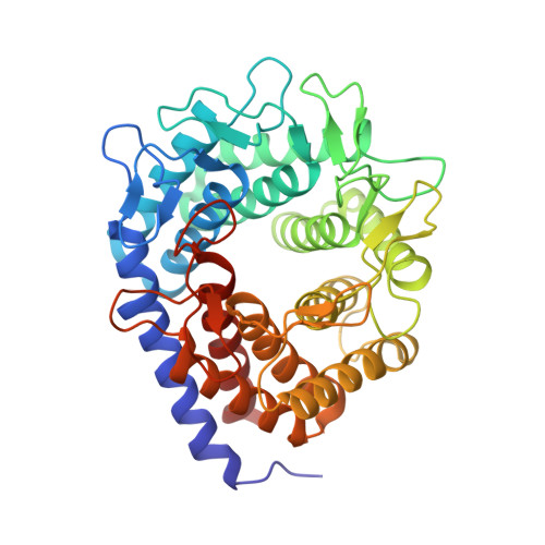
Cellobiose 2-epimerase (CE) is commonly recognized as an epimerase as most CEs mainly exhibit an epimerization activity towards disaccharides. In recent years, several CEs have been found to possess bifunctional epimerization and isomerization activities. They can convert lactose into lactulose, a high-value disaccharide that is widely used in the food and pharmaceutical industries. However, the factors that determine the catalytic direction in CEs are still not clear. In this study, the crystal structures of three newly discovered CEs, CsCE (a bifunctional CE from Caldicellulosiruptor saccharolyticus), StCE (a bifunctional CE from Spirochaeta thermophila DSM 6578) and BtCE (a monofunctional CE from Bacillus thermoamylovorans B4166), were determined at 1.54, 2.05 and 1.80 Å resolution, respectively, in order to search for structural clues to their monofunctional/bifunctional properties. A comparative analysis of the hydrogen-bond networks in the active pockets of diverse CEs, YihS and mannose isomerase suggested that the histidine corresponding to His188 in CsCE is uniquely required to catalyse isomerization. By alignment of the apo and ligand-bound structures of diverse CEs, it was found that bifunctional CEs tend to have more flexible loops and a larger entrance around the active site, and that the flexible loop 148-181 in CsCE displays obvious conformational changes during ligand binding. It was speculated that the reconstructed molecular interactions of the flexible loop during ligand binding helped to motivate the ligands to stretch in a manner beneficial for isomerization. Further site-directed mutagenesis analysis of the flexible loop in CsCE indicated that the residue composition of the flexible loop did not greatly impact epimerization but affects isomerization. In particular, V177D and I178D mutants showed a 50% and 80% increase in isomerization activity over the wild type. This study provides new information about the structural characteristics involved in the catalytic properties of CEs, which can be used to guide future molecular modifications.
Organizational Affiliation:
State Key Laboratory of Food Science and Technology, School of Food Science and Technology, Jiangnan University, Wuxi 214122, People's Republic of China.