6RVB

NADH-dependent Coenzyme A Disulfide Reductase soaked with NADH


Experimental Data Snapshot

  • Method: X-RAY DIFFRACTION
  • Resolution: 2.90 Å
  • R-Value Free: 
    0.246 (Depositor), 0.240 (DCC) 
  • R-Value Work: 
    0.201 (Depositor), 0.190 (DCC) 
  • R-Value Observed: 
    0.202 (Depositor) 

Starting Model: experimental
View more details

wwPDB Validation   3D Report Full Report


Ligand Structure Quality Assessment 

Created with Raphaël 2.3.0Worse 01 BetterLigand structure goodness of fit to experimental dataBest fitted FADClick on this verticalbar to view details

This is version 1.3 of the entry. See complete history


Literature

Characterization and X-ray structure of the NADH-dependent coenzyme A disulfide reductase from Thermus thermophilus.

Lencina, A.M.Koepke, J.Preu, J.Muenke, C.Gennis, R.B.Michel, H.Schurig-Briccio, L.A.

(2019) Biochim Biophys Acta Bioenerg 1860: 148080-148080

  • DOI: https://doi.org/10.1016/j.bbabio.2019.148080
  • Primary Citation of Related Structures:  
    6RUZ, 6RVB, 6RVH

  • PubMed Abstract: 

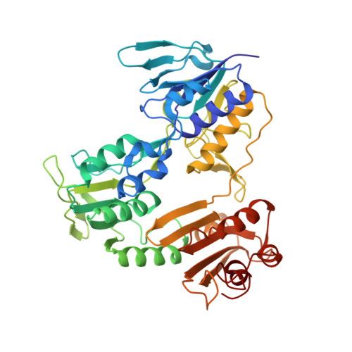
    The crystal structure of the enzyme previously characterized as a type-2 NADH:menaquinone oxidoreductase (NDH-2) from Thermus thermophilus has been solved at a resolution of 2.9 Å and revealed that this protein is, in fact, a coenzyme A-disulfide reductase (CoADR). Coenzyme A (CoASH) replaces glutathione as the major low molecular weight thiol in Thermus thermophilus and is maintained in the reduced state by this enzyme (CoADR). Although the enzyme does exhibit NADH:menadione oxidoreductase activity expected for NDH-2 enzymes, the specific activity with CoAD as an electron acceptor is about 5-fold higher than with menadione. Furthermore, the crystal structure contains coenzyme A covalently linked Cys44, a catalytic intermediate (Cys44-S-S-CoA) reduced by NADH via the FAD cofactor. Soaking the crystals with menadione shows that menadione can bind to a site near the redox active FAD, consistent with the observed NADH:menadione oxidoreductase activity. CoADRs from other species were also examined and shown to have measurable NADH:menadione oxidoreductase activity. Although a common feature of this family of enzymes, no biological relevance is proposed. The CoADR from T. thermophilus is a soluble homodimeric enzyme. Expression of the recombinant TtCoADR at high levels in E. coli results in a small fraction that co-purifies with the membrane fraction, which was used previously to isolate the enzyme wrongly identified as a membrane-bound NDH-2. It is concluded that T. thermophilus does not contain an authentic NDH-2 component in its aerobic respiratory chain.


  • Organizational Affiliation

    Department of Biochemistry, University of Illinois, 600 S. Mathews Street, Urbana, IL 61801, USA.


Macromolecules
Find similar proteins by:  (by identity cutoff)  |  3D Structure
Entity ID: 1
MoleculeChains Sequence LengthOrganismDetailsImage
NADH oxidase
A, B, C, D
443Thermus thermophilusMutation(s): 0 
Gene Names: TT_C1484
EC: 1.6
UniProt
Find proteins for Q72HK3 (Thermus thermophilus (strain ATCC BAA-163 / DSM 7039 / HB27))
Explore Q72HK3 
Go to UniProtKB:  Q72HK3
Entity Groups  
Sequence Clusters30% Identity50% Identity70% Identity90% Identity95% Identity100% Identity
UniProt GroupQ72HK3
Sequence Annotations
Expand
  • Reference Sequence
Small Molecules
Ligands 3 Unique
IDChains Name / Formula / InChI Key2D Diagram3D Interactions
FAD (Subject of Investigation/LOI)
Query on FAD

Download Ideal Coordinates CCD File 
E [auth A],
H [auth B],
K [auth C],
N [auth D]
FLAVIN-ADENINE DINUCLEOTIDE
C27 H33 N9 O15 P2
VWWQXMAJTJZDQX-UYBVJOGSSA-N
COA
Query on COA

Download Ideal Coordinates CCD File 
G [auth A],
J [auth B],
M [auth C],
P [auth D]
COENZYME A
C21 H36 N7 O16 P3 S
RGJOEKWQDUBAIZ-IBOSZNHHSA-N
NAD
Query on NAD

Download Ideal Coordinates CCD File 
F [auth A],
I [auth B],
L [auth C],
O [auth D]
NICOTINAMIDE-ADENINE-DINUCLEOTIDE
C21 H27 N7 O14 P2
BAWFJGJZGIEFAR-NNYOXOHSSA-N
Experimental Data & Validation

Experimental Data

  • Method: X-RAY DIFFRACTION
  • Resolution: 2.90 Å
  • R-Value Free:  0.246 (Depositor), 0.240 (DCC) 
  • R-Value Work:  0.201 (Depositor), 0.190 (DCC) 
  • R-Value Observed: 0.202 (Depositor) 
Space Group: P 42 21 2
Unit Cell:
Length ( Å )Angle ( ˚ )
a = 160.75α = 90
b = 160.75β = 90
c = 256.92γ = 90
Software Package:
Software NamePurpose
PHENIXrefinement
XDSdata reduction
XSCALEdata scaling
PHASERphasing

Structure Validation

View Full Validation Report


Ligand Structure Quality Assessment 

Created with Raphaël 2.3.0Worse 01 BetterLigand structure goodness of fit to experimental dataBest fitted FADClick on this verticalbar to view details

Entry History 

Deposition Data

Revision History  (Full details and data files)

  • Version 1.0: 2019-09-25
    Type: Initial release
  • Version 1.1: 2019-10-23
    Changes: Data collection, Database references
  • Version 1.2: 2024-01-24
    Changes: Data collection, Database references, Refinement description
  • Version 1.3: 2024-11-13
    Changes: Structure summary